Perinium (Viva)
6 years ago 3019
Q.1 What are the boundaries of the perineum?
Superficial :
Anterior:
Scrotum in male
Mons pubis in female
Posterior: Buttocks.
Lateral:
The upper part of the medial side of the thigh
Deep:
Anterior:
The upper part of the pubic arch
Arcuate pubic ligament.
Posterior: Tip of the coccyx.
Lateral:
Conjoined ischiopubic rami,
Ischial tuberosity, and
Sacrotuberous ligament
Q.2 What are the divisions of the perineum?
An imaginary transverse line joining the anterior parts of ischial tuberosities divides the rhomboid-shaped perineum into two triangular regions:
Q.3 What are the boundaries of the urogenital triangle?
Q.4 What are the boundaries of the anal triangle?
Q.5 What is the perineal body?
The fibromuscular structure in the median plane is about 1.25 cm in front of the anal margin. Supports pelvic organs in females.
Q.6 Name the muscles forming the perineal body.
Nine muscles:
Q.7 What is the clinical importance of the perineal body?
In females, it may rupture during childbirth, which if unrepaired may lead to prolapse of the urinary bladder, uterus, and rectum.
Q.8 What are the boundaries of the ischiorectal fossa?
It is a wedge-shaped space, on each side of the anal canal below the pelvic diaphragm between the obturator internus and levator ani.
| Base: Skin |
| Apex: Meeting of obturator fascia with an inferior layer of pelvic fascia. |
| Anterior: The posterior border of perineal membrane. |
| Posterior: Lower border or gluteus maximus and sacrotuberous ligament. |
| Lateral wall: Obturator internus with fascia, Medial surface of ischial tuberosity. |
| Medial wall: External anal sphincter, in the lower part, Levator ani fascia, in upper part. |
Q.9 What are the contents of the ischiorectal fossa?
Q.10 Why the infections of perianal space are very painful but those of ischiorectal space are much less painful?
Fat in perianal space is tightly arranged in small loculi formed by complete septa therefore little swelling due to infections causes increased tension and pain, but in ischiorectal space, fat is loosely arranged therefore swelling can occur without tension.
Q.11 Why infections are more common in the ischiorectal fossa?
Because of the presence of poorly vascularized fat in the fossa, this region is very vulnerable to infection. Infections usually reach the fossa from the anal canal.
Q.12 Why the unilateral ischiorectal abscess if not drained becomes bilateral?
Because of the extension of infection through the horse-shoe recess behind the anal canal which connects the fossa of both sides.
Q.13 What is Hiatus of Schwalbe?
This is the gap between the obturator fascia and the origin of the levator ani. Herniation of some pelvic contents can take place through the gap.
Q.14 Why in debilitating disorders prolapse of the rectum occurs?
Because of:
Q.15 Why the abscesses of the ischiorectal fossa can be drained by incision easily?
Q.16 Name the structures passing through the gap between the arcuate pubic and transverse perineal ligament.
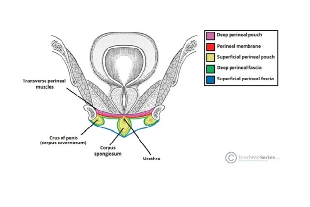
Q.17 How the deep perineal space is formed?
The deep perineal space is formed between the superior and inferior fascia of the urogenital diaphragm.
Q.18 What are the contents of deep perineal space in males?
Q.19 What are the contents of superficial perineal space in males?
Q.20 Name the structures piercing the perineal membrane (inferior fascia of the urogenital diaphragm).
In males:
In females:
Q.21 Name the structures forming the urogenital diaphragm.
Q.22 Name the female external genital organs.
Q.23 What are the boundaries of the gynecological perineum?
The area between the posterior commissure (skin connecting prominent posterior ends of labia majora) and anus, constitutes the gynecological perineum.
Q.24 What is the position of glands of Bartholin?
These are homologous with bulbourethral glands (of Cowper) in males. Lie in the superficial perineal space at the vaginal orifice. The duct of each gland opens at the side of the hymen, between the hymen and labium minor.
Q.25 How pudendal canal is formed?
Q.26 What are the contents of the pudendal canal (Alcock’s canal)?
Q.27 What are the structures supplied by the pudendal nerve?
Q.28 Where is the ‘pudendal nerve block’ given in vaginal operations?
Near the ischial spine, a needle passed through the vaginal wall and then guided by a finger.
Q.29 What are the branches of the internal pudendal artery?
Comments (0)