Vagina (Viva)
6 years ago 3012
Q.1 What is the position and extent of the vagina?
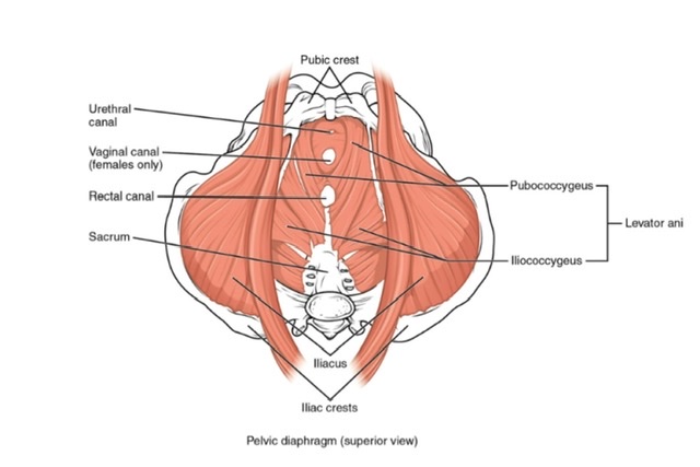
It is situated behind the bladder and urethra and in front of the rectum and anal canal. It extends from the vulva to the uterus.
Q.2 What are the variations in the shape of the lumen of the vagina?
Q.3 What are the relations of the vagina?
Anterior wall: 8 cm long
Posterior wall: 10 cm long
Lateral wall: On each side
Q.4 What are the fornices of the vagina?
The upper part of the vagina is converted into a circular groove by protruding cervix, which is divided into four parts known as vaginal fornices: anterior, posterior and two lateral fornices. The anterior fornix is the shallowest and the posterior fornix deepest.
Q.5 What is the arterial supply of the vagina?
Q.6 What is the lymphatic drainage of the vagina?
Q.7 What is the nerve supply of the vagina?
Q.8 What is the characteristic feature of the lining epithelium of the vagina?
The vagina is lined by stratified squamous epithelium and has no glands. It is lubricated partly by cervical mucus and partly by desquamated vaginal epithelial cells.
Q.9 What important information can be obtained by per vaginal (PV) examination?
The condition of:
• Vagina:
Abnormalities of entrance or walls.
• Urethra:
Can be rolled against symphysis in the anterior wall.
• Rectum:
If it contains a tumor, foreign body, or feces.
• Cervix and external os
• Vaginal fornices
• Rectovaginal pouch:
By finger in the posterior fornix. If it contains uterus, prolapsed ovaries, tumors, or abdominal collection of fluid.
• Ureter:
If thickened or has a stone, can be rolled against the pelvic bone just before it enters the bladder.
• To determine the diagonal conjugate of the pelvis, to assess whether the pelvis is large enough to transmit fetal head.
Q.10 What is a hymen?
A fold of mucous membrane at the lower end of the vagina which partially closes it.
Q.11 What is the developmental origin of the vagina?
The vagina is formed by the development of the lumen within the vaginal plate, which is formed by:
Q.12 What are the abnormalities of the vagina?
Comments (0)