Uterus (Viva)
6 years ago 3024
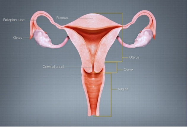
Q.1 What are the parts of the uterus?
Q.2 What are the parts of the cervix?
Q.3 What are ‘arbor vitae’?
The mucous membrane of the cervical canal is thrown into the fold and oblique furrows which pass away from anterior and posterior vertical ridges.
Q.4 What are the angulations of the uterus?
Q.5 Name the structures attached to the lateral border of the body of the uterus.
Q.6 What are the supports of the uterus?
The uterus is prevented from sagging down by a number of factors. They are classified into :
Q.7 How uterine axis support the uterus in maintaining its position?
The anteversion prevents the uterus from sagging through the vagina. Any rise in intraabdominal pressure tends to push uterus against bladder, which further accentuates anteversion. The angle of anteversion is maintained by uterosacral and round ligaments.
Q.8 What is the canal of Nuck?
The round ligament of the uterus in the inguinal canal, in fetal life, is accompanied by a process of the peritoneum, which if persists, after birth is known as the canal of Nuck.
Q.9 What are the parts of the broad ligament of the uterus?
Q.10 What are the contents of the broad ligament of the uterus?
Q.11 What is the arterial supply of the uterus?
Q.12 Name the structures supplied by the uterine artery.
Q.13 What is the histological structure of the uterus?
The wall of the uterus is made up of 3 layers:
Q.14 What is the histological difference between the two parts of the cervix?
The vaginal portion is covered by squamous epithelium which becomes continuous with columnar cells of the cervical canal at external os.
Q.15 Describe the course of the uterine artery and its distribution to the uterus.
The uterine artery is a branch of the anterior trunk of the internal iliac artery.
It runs downwards and forward and when reaches the parametrium, it turns medially towards the uterus. It reaches the uterus at the level of the internal os, where it turns upwards at right angles and runs a spiral course along the lateral border of the uterus to the uterine cornu.
During the vertical part, it gives branches, which run transversely into the myometrium (Arcuate arteries). From these arise radial arteries at right angles and they reach basal layers of the endometrium (Basal arteries). Basal arteries give rise to terminal spiral and straight arterioles of the endometrium.
Q.16 What is the lymphatic drainage of the uterus?
Lymphatics of the uterus form three intercommunicating plexuses, which drain into:
Q.17 What are the changes in uterus with age?
| In fetal life: The cervix is larger than the body of the uterus. |
| At puberty: Uterus enlarges. The body grows more than the cervix so it acquires its pyriform shape. |
| During menstruation: Uterus slightly enlarged and more vascular. |
| During pregnancy: Uterus enormously enlarged. |
| After pregnancy: Regresses to normal size but the thickness of the wall and size of the cavity remains larger. |
| In older age: The uterus is smaller and denser in texture. |
Q.18 How is the uterus divided into upper and lower segments during pregnancy?
The uterus is divided into the upper uterine segment consisting of the fundus and the greater part of the body and the lower segment consisting of the lower part of the body and cervix. The upper one-third of the cervix is known as the isthmus.
Q.19 What is the developmental origin of the uterus?
The epithelium of the uterus develops from fused paramesonephric ducts. Myometrium from surrounding mesoderm. The unfused part of the paramesonephric duct embedded in the myometrium forms the fundus.
Q.20 What are the common anomalies of the uterus?
Q.21 What are the advantages and disadvantages of a midline incision made in the uterus?
The midline part of the uterus is the least vascular part, so there is less bleeding during surgery but the wound also heals poorly due to poor vascularity.
Q.22 What precaution should be taken in relation to the ureter while removing the uterus (hysterectomy)?
At the supravaginal cervix, the ureter lies just above the level of the lateral fornix and below uterine vessels as these pass within the broad ligament. In hysterectomy, the ureter may be accidentally divided when clamping the uterine vessels, especially when pelvic anatomy is distorted.
Q.23 What is the fate of mesonephric ducts and tubules in females?
They form a number of vestigial structures.
Q.24 How vesicular appendix is developed?
From the cranial part of the paramesonephric duct
Comments (0)