Ovary & Uterine Tube (Viva)
OVARY
Q.1 What is the position of the ovary?
It lies in the ovarian fossa on the lateral pelvic wall, just below and behind the lateral part of the uterine tube.
Q.2 What are the boundaries of the ovarian fossa?
Inferior: Obliterated umbilical artery.
Posterior: Ureter and internal iliac vessels
Anterior: External iliac vessels.
Q.3 What are the peritoneal relations of ovary?
- Ovary is entirely covered with peritoneum except along the anterior border where the two layers of peritoneum are continuous with the posterior layers of the broad ligament of uterus, as mesovarium.
- Lateral part of the broad ligament, from infundibulum of tube and upper pole of the ovary to external iliac vessels, forms suspensory ligament of the ovary.
Q.4 What is the blood supply of ovary?
- Arterial supply:
– Ovarian artery.
– Branches of the uterine artery.
- Venous drainage:
Pampiniform plexus which condense into a single vein, near the pelvic inlet.
Right ovarian vein, drains into inferior vena cava. Left ovarian vein, drains into the left renal vein.
Q.5 What is the histological structure of ovary?
Ovary is made up of (from within outwards):
- Medulla:
Vascular connective tissue having vessels, nerves, and lymphatics.
- Cortex:
Has various stages of development of ovarian follicles.
- Tunica albuginea: T
hin layers of connective tissue.
- Germinal epithelium:
Made up of cuboidal cells, derived from the peritoneum.
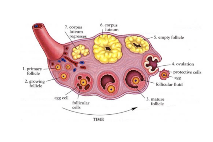
Q.6 What is ovulation? What are its indicates?
Release of one or more ova from one of the ovaries during each menstrual cycle is termed as ovulation. Development of an ovum followed by the process of ovulation.
UTERINE TUBES (FALLOPIAN TUBES)
Q.1 What are the parts of the uterine tube?
- Infundibulum (Fimbriated):
Opens into peritoneal cavity by abdominal ostium.
- Ampulla:
Forms lateral 2/3 of tube.
Thin-walled and wider lumen.
- Isthmus:
Forms medial 1/3 tube.
Thick-walled and narrow lumen.
- Uterine (Interstitial):
Lies within the uterine wall and opens into the uterine cavity by uterine ostium.
Q.2 What are the relations of the uterine tube with ovary?
- Near lateral pelvic wall, ampulla is related to anterior and posterior borders, upper pole, and medial surface of ovary.
- One of the fimbria is long and is attached to the tubal (upper) pole of the ovary, it is known as ovarian fimbria.
Q.3 What is mesosalphinx?
It is part of the broad ligament between mesovarium and uterine tube.
Q.4 What is the blood supply of the uterine tube?
Arterial supply:
- Medial 2/3: Uterine artery.
- Lateral 1/3: Ovarian artery.
Venous drainage:
Into pampiniform plexus and uterine veins.
Q.5 What is the histological structure of uterine tubes?
They are made up of the following coats:
- Outer: Serous coat
- Middle: Muscular coat has circular muscles.
- Inner: Mucous membrane is lined by ciliated columnar cells and nonciliated secretory cells.
The mucous membrane forms folds which fill up the lumen of the tube.
Q.6 How uterine tubes are developed?
From unfused parts of paramesonephric ducts. The point of the invagination of the duct remains as the abdominal openings.
Q.7 At what site the fertilization of ovum takes place?
In the ampulla of the fallopian tube
Q.8 What is tubectomy?
Female sterilization, in which 2 to 3 cm long segment of the tube is excised and cut ends are ligated.
Comments (0)