Male External Genital Organs (Viva)
6 years ago 2487
Q.1 What are the parts of the penis?
Q.2 What is Buck’s fascia?
It is the membranous layer of superficial fascia of the penis.
Q.3 What is the arterial supply of the penis?
Q.4 What is the lymphatic drainage of the penis?
The glans penis drains into deep inguinal nodes and rest of the penis into the upper medial group of superficial inguinal lymph nodes.
Q.5 What is the developmental origin of the penis?
The genital tubercle at the cranial end of the cloacal membrane, which lengthens to form phallus which enlarges to form the penis.
Q.6 What is scrotum?
It is a cutaneous bag containing testis, epididymis, and lower part of spermatic cord.
Q.7 Name the structures forming layers of scrotum. From without inwards:
Q.8 What is the blood supply of scrotum.
Q.9 What is the nerve supply of scrotum?
Q.10 Why the extravasation of fluid into the scrotal sac is bilateral?
Because the septum which divides scrotum into right and left compartments, is incomplete superiorly.
Q.11 What is the situation of testis?
Q.12 What is sinus of epididymis?
It is the extension of the cavity of tunica vaginalis between testis and epididymis from its lateral side, on the posterior border.
Q.13 What is the Appendix of testis?
Q.14 What are the coverings of testis?
From without inwards:
Q.15 What is the arterial supply of testis?
Testicular artery: Branch of abdominal aorta.
At the posterior border of the testis, it divides into branches:
Q.16 What is pampiniform plexus?
Ultimately, one vein is formed which drains into inferior vena cava on the right side and left renal vein on the left side.
Q.17 What is the lymphatic drainage of testis?
Pre-aortic and para-aortic lymph nodes at the level of L2 vertebra.
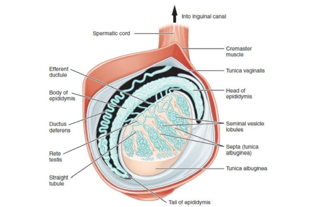
Q.18 What is the structure of the testis?
Testis is divided into 200-300 lobules by septae passing from mediastinum testis to tunica albuginea each containing one to three seminiferous tubules. The tubules anastomose posteriorly into 20-30 straight tubules. These unite in mediastinum testis to form, Rete testis from which efferent ducts arise and pass into the head of the epididymis.
Q.19 What is the developmental origin of testis?
Testis arises from mesodermal genital ridge in the posterior abdominal wall just medial to developing kidney and links up with epididymis and vas, which develop from mesonephric duct (Wolffian duct).
Q.20 What is Gubernaculum testis?
It is a fibromuscular band attaching the testis to the bottom of the scrotum. According to Hunter, gubernaculum forms the inguinal canal by its passage through the abdominal wall. It develops from a mesenchymal strand.
Q.21 What is processus vaginalis?
It is the prolongation of the peritoneal cavity projecting into the scrotum. The testis in scrotum slides posterior to this and projects into it. Thus the testis is covered by peritoneum from front and sides. About the time of birth, it obliterates, leaving the testis covered by tunica vaginalis.
Q.22 What are the positions of testis during its descent in fetal life?
• 3rd month: Reaches iliac fossa
• 7th month: Deep inguinal ring
• During 7th month: Traverses inguinal canal
• 8th month: Reaches superficial inguinal ring
• Beginning of 9th month; Descends into the scrotum.
Q.23 Why the cervical lymph nodes become enlarged in tumors arising from testis?
Because of the plentiful communications of para-aortic lymph nodes in abdomen with those of the thoracic region and which in turn communicate with cervical nodes.
Q.24 What is varicocele?
It is the dilatation of pampiniform plexus of veins. It is commoner on the left side because of left testicular veins compression by a loaded sigmoid colon, left kidney tumor which invades renal veins and obstructs the drainage of left testicular veins, obstruction by angulation at the site of entry of left testicular veins into the left renal vein, in which pressure is higher than in inferior vena cava.
Q.25 What are ectopic testis?
The testis descends but is found in an unusual position, e.g. under the skin of the front of the abdomen, under the skin of thigh in the femoral canal, under the skin of the penis, or in perineum behind the scrotum.
In these cases, the cord is long (unlike the undescended testes).
Q.26 What is a hydrocele? What are the two different types of hydrocele?
Hydrocele occurs due to the accumulation of fluid within tunica vaginalis of the scrotum or along the spermatic cord.
Hydroceles can be of two types:
Communicating and non-communicating hydroceles
Communicating hydrocele occurs due to incomplete closure of tunica vaginalis. As a result, there is a communication with the fluids of the abdominal cavity. As a result, there may be continuous variation in the size of the hydrocele. This type of hydrocele is usually present at birth.
Non-communicating hydrocele:
This type of hydrocele may be present at birth or develop years later for no obvious reason. It usually remains the same in size or has a very slow rate of growth. The pathophysiology of hydrocele is related to either increased fluid production or impaired this absorption.
Q.27 What is Monorchidism?
Developmental absence of a testicle.
Q.28 What is vas aberrans of Haller?
It is a blind tube that lies between the tail of the epididymis and the commencement of vas.
Q.29 What is the length of epididymis?
When uncoiled 20 feet, but in coiled form, the comma-shaped body is only 1 inch long on the posterolateral aspect of testis.
Q.30 What is the developmental origin of the Appendix of epididymis?
Represents cranial end of the mesonephric duct. Also known as pedunculated hydatid of Morgagni.
Q.31 What is organ of Giraldes (Paradidymis)?
Free tubules in spermatic cord above head of epididymis.
Represent caudal mesonephric tubules.
Q.32 What is the extent of the spermatic cord?
It extends from the upper pole of the testis, through the inguinal canal to the deep inguinal ring.
Q.32.1 What are the coverings of the spermatic cord?
From within outwards
Q.33 What are the constituents of the spermatic cord?
Comments (0)