Ureter (Viva)
6 years ago 4875
Q.1 What are ureters?
These are pair of narrow, thick-walled muscular tubes which convey urine from the kidneys to the urinary bladder.
Q.2 What is the length of the ureter?
Each ureter is about 25 cm long, of which the upper half lies in the abdomen and the lower half in the pelvis.
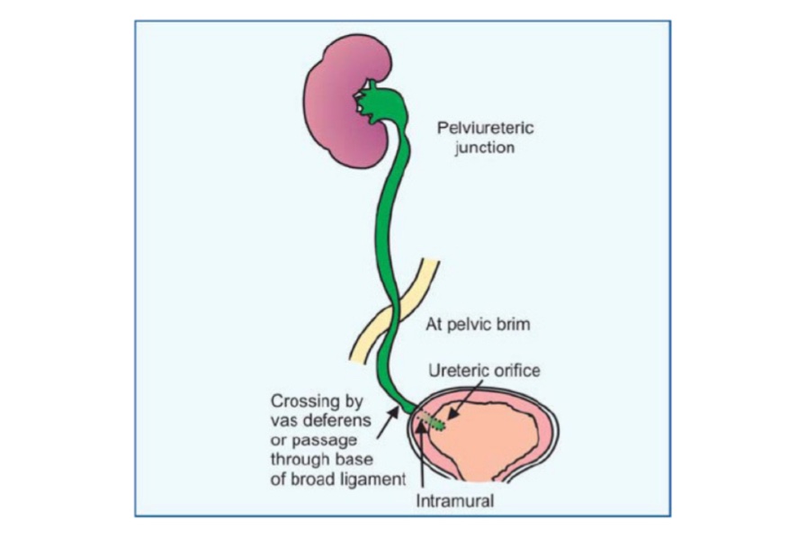
Q.3 What is the course in the ureter?
The course of ureter is divided into two parts :
Ureter begins at the renal pelvis (Funnel-shaped dilatation) from the hilus of the kidney and descends along its medial border. It gradually narrows and becomes ureter proper at the lower pole of the kidney. It descends on psoas muscle and enters pelvis by crossing in front of the termination of common iliac artery
It first runs downwards, backward and laterally, following the anterior margin of the greater sciatic notch. Opposite ischial spine it turns forwards and medially to reach the base of the urinary bladder. Ureter enters the bladder wall obliquely and opens at the lateral angle of the trigone. Its point of termination corresponds to the pubic tubercle.
Q.4 Name the sites at which constrictions are present in the ureter.
Three sites:
Q.5 What is the clinical importance of constrictions of ureter?
A ureteric calculus is likely to lodge at one of these three levels as described in
Q.6 What are structures crossing the abdominal part of the right ureter from medial to lateral side?
Q.7 What are the structures crossing the abdominal part of the left ureter from medial to lateral side?
Q.8 What are the relations of the ureter in its forward course in pelvis?
In males:
In females:
Q.9 What is the arterial supply of ureter?
Q.10 What is the nerve supply of ureter?
They reach through renal, aortic, and both hypogastric plexus.
Autonomic nerves to ureter are predominantly sensory in function.
Q.11 What is renal colic?
It is spasm of the ureter by a stone. There is a sudden, agonizing pain in the loin.
Q.12 Where the pain of renal colic is referred?
Pain is referred to cutaneous area innervated by T11-L2.
Q.13 What is the developmental origin of ureter?
From part of ureteric bud that lies between the pelvis of the kidney and vesico-urethral canal.
Q.14 What are the congenital abnormalities of ureter?
Q.15 What is ‘post caval’ ureter? What is its clinical importance?
The right ureter instead of lying to the right of inferior vena cava may pass behind it.
Clinical importance:
May lead to compression of ureter and obstruction to the flow of urine.
Q.16 How the reflux of urine from the bladder into ureter is prevented?
The intravesical oblique course of the ureter has valvular action which prevents the reflux of urine from bladder to ureter.
Comments (0)