Kidney & Suprarenal Glands (Viva)
6 years ago 3629
KIDNEYS
Q.1 Where are the kidneys situated?
The kidneys are situated retroperitoneally on the posterior abdominal wall on each side of the vertebral column. The right kidney is slightly lower than the left and the left kidney is a little nearer to the median plane.
Q.2 What is the extent of the kidney in relation to the vertebral column?
The kidneys vertically extend from the upper border of the T12 vertebra to the centre of the body of the L3 vertebra. The right kidney is slightly lower than the left.
Q.3 What is the relation of the transpyloric plane to kidneys?
Transpyloric plane passes through the upper part of the hilus of the right kidney and through the lower part of the hilus of the left.
Q.4 What are the measurements of the normal kidney?
Left kidney is a little longer and narrower.
Q.5 What are the anterior relations of the kidneys?
Right kidney:
Right suprarenal
Liver
Second part of duodenum
Hepatic flexure of colon
Small intestine
Hepatic and intestine surfaces are covered by peritoneum
Left kidney:
Left suprarenal
Stomach
Spleen
Pancreas
Jejunum
Splenic flexure
Descending colon and splenic vessels.
The gastric, splenic, and jejunal surfaces are covered by peritoneum.
Q.6 What are the posterior relations of the kidney?
Both kidneys are related to:
The right kidney is also related to the 12th rib and the left kidney to 11th and 12th ribs.
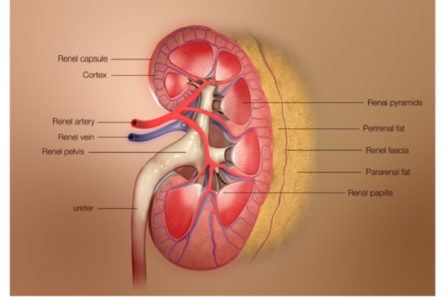
Q.7 What are the coverings of the kidneys?
From within outwards the coverings are:
Superiorly, two layers of renal fascia first enclose the suprarenal gland in a separate compartment, then they fuse with each other and become continuous with the fascia on the undersurface of diaphragm.
Inferiorly, the two layers remain separate and enclose ureter. Laterally, the two layers fuse and become continuous with fascia transversalis.
Medially, the anterior layer passes in front of renal vessels and fuses with connective tissue around the aorta and inferior vena cava. The posterior layer fuses with fascia covering quadratus lumborum and psoas major. At the medial border of kidney fascia forms a septum.
Q.8 What is the Fascia of Toldt and Fascia of Zuckerkandl?
The anterior layer of renal fascia is known as the fascia of the Toldt and posterior layer as the fascia of Zuckerkandl.
Q.9 What are the structures found at the hilus of the kidney?
From before backward:
Q.10 What are the vascular segments of the kidney?
Each renal artery at the hilus of the kidney divides into an anterior and posterior branch, which in turn divides into segmental arteries that supply a definite part (segment) of the kidney. In each kidney, there are five segments, i.e. apical, upper, lower, middle, and posterior.
Q.11 What is the clinical importance of vascular segments of the kidney?
Each segmental artery is an end artery, so the vascular segments are independent units. So the intersegmental incisions are given for the removal of a part of the kidney.
Q.12 What is the direction of blood flow in ruptured kidney or pus in perinephric abscess?
First, it causes distension of renal fascia and then downwards into pelvis within the fascial compartment. The mid-line attachment of renal fascia and fascial septum prevents extravasation to the opposite side.
Q.13 What care should be taken in the exposure of kidneys from behind when the 12th rib is to be excised?
Push up the pleura which crosses the medial half of the 12th rib.
Q.14 How the kidney are developed?
Kidneys are formed in the sacral region and then ascend upwards. The kidney develop from:
Q.15 Name the common congenital abnormalities of kidneys.
SUPRARENAL (ADRENAL) GLANDS
Q.1 What is the position of adrenal glands?
Q.2 What are the parts of adrenal glands seen in cross-section?
The volume of medulla is about one-tenth of the cortex.
Q.3 What is chromaffin system?
Q.4 What are the components of the chromaffin system?
Q.5 What are the different layers of the adrenal cortex?
Q.6 What is the blood supply of suprarenal glands?
Arterial supply:
Venous drainage:
Q.7 Name of structures lying between two suprarenal glands.
Q.8 Compare the two suprarenal glands.
| Left | Right | |
| Shape | Semilunar | Triangular |
| Size | Larger | Smaller |
| Position | Upper part of medial border of kidney | Upper part of the anterior surface of kidney |
| Level | Lower | Higher |
| Hilum | Near lower end | Near upper end |
| Peritoneal | Separated from stomach by peritoneum | Only lower part relations related to peritoneum |
| Visceral relations: | ||
| Anterior surface | Superior: Stomach Inferior: Pancreas Splenic artery |
Medial: Inferior vena cava Lateral: Part of bare area of the liver |
| Posterior | Medial: Crus of surface Lateral: Kidney |
Inferior: Kidney, diaphragm Superior: Crus of diaphragm |
| Medial border | Left coeliac ganglion, left inferior phrenic artery, left gastric artery | Right coeliac ganglion, right inferior phrenic artery |
Comments (0)