Pancreas (Viva)
6 years ago 3550
Q.1 Why pancreas is called a ‘double gland’?
The pancreas is called a double gland because it is partly exocrine and partly endocrine.
Q.2 What are the secretions of the pancreas?
The exocrine part secretes pancreatic juice which has digestive functions. Endocrine part secretes hormones, e.g. insulin, glucagon, etc.
Q.3 At what level the pancreas lie?
The pancreas lies across the posterior abdominal wall at the level of L1 and L2 vertebra.
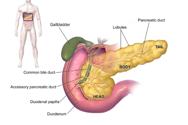
Q.4 What is the shape and different parts of the pancreas?
Pancreas is a J-shaped organ. It is divided into 4 parts:
Q.5 What are the relations of the head of the pancreas?
Anterior surface:
Posterior surface:
Superior border:
Inferior border:
Right lateral border:
Uncinate process is related anteriorly to superior mesenteric vessels and posteriorly to the aorta.
Q.6 What are the relations of the neck of the pancreas?
Anterior surface:
Posterior surface:
Q.7 What are the relations of the body of the pancreas?
Anterior surface:
Posterior surface:
Inferior surface:
Inferior border:
Superior border:
Anterior border:
Q.8 What are relations to the tail of the pancreas?
Tail of pancreas lies in lienorenal ligament and is related to the gastric surface of the spleen.
Q.9 What is the arterial supply of the pancreas?
Q.10 What is the venous drainage of the pancreas?
The pancreas drains into splenic, superior mesenteric and portal veins.
Q.11 What are the ducts draining the secretions of the exocrine part of the pancreas?
The two ducts carrying the exocrine secretion of pancreas are:
Q.12 What is ‘Pseudopancreatic cyst’?
Anterior to the pancreas lies the stomach, separated from it by the lesser sac. The sac may be closed off and distended with fluid either from perforation of posterior gastric ulcer or as a result of acute pancreatitis, thus forming a pseudo pancreatic cyst.
Q.13 Why carcinoma of head of pancreas is associated with obstructive jaundice?
The head of the pancreas lies in the C-curve of the duodenum in relation to the opening of the common bile duct. Therefore, carcinoma of the head of the pancreas will cause the compression of the common bile duct and causes obstructive jaundice.
Q.14 What is the developmental origin of the pancreas?
The ventral pouch rotates posteriorly to fuse with lower aspect of dorsal diverticulum, trapping the superior mesenteric vessels between two parts.
Q.15 What are the common developmental anomalies of the pancreas?
Q.16 How are secretions of the pancreas passed into the duodenum?
The pancreatic secretions are poured into the duodenum with the help of two ducts:
—This begins in the tail of the pancreas and passes to the right through the body. At the neck of the pancreas, it turns downwards and backward and joins the bile duct just outside the duodenal wall. The walls of the bile and main pancreatic ducts join each other here, but their lumens remain separate as the ducts descend through the muscle wall and submucosa of the duodenum.
This begins in the lower part of the head of the pancreas. It runs upwards crossing in front of the main duct and opens into the duodenum at the minor duodenal papilla (which has a short distance above and in front of the major papilla.
Comments (0)