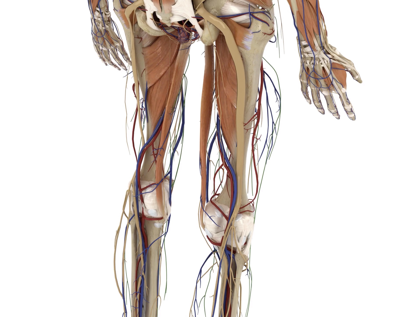
Nerves of Lower Limb (Viva)
LUMBAR PLEXUS
Q.1 How lumbar plexus is formed?
By the ventral rami L1-3 and greater part of the ventral ramus of L4. The first lumbar nerve also receives a branch from the T12 nerve.
Q.2 What are the branches of lumbar plexus?
- Muscular: – To quadratus lumborum (T12, L1-3) – Psoas minor (L1) – Psoas major (L2,3) – Iliacus (L2,3)
- Iliohypogastric nerve (L1)
- Ilioinguinal nerve (L1)
- Genitofemoral nerve (L1,2)
- Lateral cutaneous nerve of thigh (Dorsal division of ventral primary rami of L2,3)
- Femoral nerve (Dorsal division of ventral primary rami of L2-4)
- Obturator (Ventral division of ventral primary rami of L2-4)
- Accessory obturator (Ventral division of ventral primary rami of L3,4).
Q.3 What is the distribution of the obturator nerve?
- Anterior branch supplies:
– Muscular branches: To adductor longus, gracilis, obturator externus, and occasionally adductor brevis and pectineus.
– Articular: To hip joint.
– Cutaneous: To subsartorial plexus
–. Vascular branches: To femoral artery
- Posterior branch supplies:
– Muscular branches: To obturator externus, adductor magnus, and adductor brevis.
– Articular: To knee joint.
Q.4 Name the branches of the femoral nerve.
- Anterior division supplies:
– Nerve to pectineus
– Intermediate cutaneous nerve of thigh
– Medial cutaneous nerve of thigh
– Nerve to sartorius
– Nerve to iliacus
- Posterior division supplies:
– Saphenous nerve
– Muscular branches to quadriceps femoris
– Vascular branches to femoral artery
– Articular branches to hip and knee joint
Q.5 Name the nerves forming the subsatorial plexus.
- Medial cutaneous nerve of thigh
- Saphenous nerve
- Cutaneous branch of the anterior division of the obturator nerve.
Q.6 Name the nerves forming the patellar plexus.
- Saphenous nerve
- Medial, intermediate and lateral cutaneous nerve of thigh
- Saphenous nerve and its infrapatellar branch
Q.7 What is ‘meralgia paresthetica’?
It is a clinical condition characterized by pain, tingling, numbness, or anesthesia in the area of distribution of the lateral cutaneous nerve of the thigh. This nerve (a branch of the lumbar plexus) usually enters the thigh, passing deep to the inguinal ligament. Occasionally, the nerve pierces the ligament and may then be compressed by it with resultant irritation of the nerve.
Q.8 How can the pain of the adductor spasm be relieved?
By division of the obturator nerve.
Q.9 Why does a patient sometimes complain of pain in the knee when the disease is actually in the hip joint?
This is referred pain because both the hip and knee joints are supplied by the same nerves, i.e. the femoral and obturator nerves.
SACRAL PLEXUS
Q.1 How sacral plexus is formed?
By ventral primary rami of L4,5 S1-4.
Q.2 What are the branches of sacral plexus?
- Sciatic nerve (L4,5 S1-3)
- Superior gluteal nerve (Posterior division of L4,5 S1)
- Inferior gluteal nerve (Posterior division of L5, S1,2)
- Perforating cutaneous nerve (Posterior division of S2,3)
- Nerve to piriformis (Posterior division of S1,2)
- Pudendal nerve (Anterior division of S1- 3)
- Posterior cutaneous nerve of thigh (Anterior division of S1,2 and posterior division of S2,3)
- Nerve to obturator internus (Anterior division of L5, S1,2)
- Nerve to quadratus femoris (Anterior division fo L4,5 S1)
- Nerve to levator ani and coccygeus and sphincter ani externus from S4 branches
- Pelvic splanchnic nerve from S2-4
Q.3 How the sciatic nerve is formed? What are its branches?
The sciatic nerve is the continuation of the sacral plexus and derives its fibers from the L4,5, S1, 2, 3. It is the largest nerve in the body. The main trunk of the sciatic nerve is the nerve of the flexor compartment of the thigh.
Branches:
- Articular: To hip joint.
- Muscular: To biceps femoris, semitendinosus, semimembranosus, and ischial head of adductor magnus.
- Terminal:
– The tibial nerve is the nerve of the flexor compartments of the thigh (through the parent trunk), leg and sole of the foot. It receives fibers from the anterior divisions of L4,5 S1,2 and S3 (which does not divide into anterior and posterior division)
– The common peroneal nerve is the nerve of the extensor and peroneal compartments of the leg and dorsum of the foot. It is derived from the posterior divisions of L4,5 S1, 2.
Q.3. Give the surface marking of the sciatic nerve.
- The sciatic nerve is represented by a thick line (2 cm broad) joining the following three points.
- The first point is taken 2.5 cm lateral to the mid-point of a line joining the posterior superior iliac spine (marked by a dimple lateral to the natal cleft) and the ischial tuberosity.
- The second point is taken at the mid-point between the greater trochanter of the femur and the ischial tuberosity.
- The third point is taken at the mid-point of a transverse line drawn at the junction of the middle and lower 2/3 of the back of the thigh, i.e. apex of the popliteal fossa.
Q.4 What will be the effect of a complete lesion of the sciatic nerve in the gluteal region?
- Motor loss:
– Loss of flexion of the knee due to paralysis of the hamstring muscles, but some weak movement is possible due to the action of the sartorius (femoral nerve) and gracilis (obturator nerve).
– Loss of all movements below the knee due to paralysis of all the muscles of the leg and foot. There will be a ‘foot drop’ deformity.
– Loss of Achilles jerk and plantar reflex.
- Sensory loss: On the outer side of the leg and almost the entire foot.
Q.5 What is ‘sciatica’ and what is its common cause?
Sciatica is the term applied when pain is felt along the course and distribution of the sciatic nerve, i.e., in the buttock, posterior aspect of the thigh and leg, and lateral aspect of the leg and foot. This is due to irritation of one or more of the roots of the sciatic nerve and commonly occurs due to a prolapsed intervertebral disc in the lumbar region.
Q.6 At what site intramuscular injections are given in the gluteal region?
The injections are given in the upper and outer quadrant of the gluteal region to avoid injury to the sciatic nerve.
Q.7 What is the site for the local anesthetic to be injected for sciatica to relieve the pain?
The site of injection is midway between the greater trochanter of the femur and the ischial tuberosity.
Q.8 What are the branches of common peroneal nerve?
- Lateral cutaneous nerve of calf
- Communicating branch to sural nerve
- Terminal branches: Deep and superficial peroneal nerve.
Q.9 Where is the common peroneal (lateral popliteal) nerve commonly injured and what are the common causes of the injury?
The nerve is commonly injured where it winds around the neck of the fibula. It may be damaged at this site by the pressure of a tight bandage of plaster cast, in severe adduction injury to the knee, or from direct trauma.
Q.10 What will be the effects of a complete section of the common peroneal (lateral popliteal) nerve at the level of the neck of the fibula?
- Motor loss:
– Inability to extend the foot or toes due to paralysis of the ankle and foot extensors (tibialis anterior, extensor hallucis longus, extensor digitorum longus, peroneus tertius and extensor digitorum brevis). This results in “foot drop” which is characteristic of the common peroneal nerve injury.
– Inability to evert the foot due to paralysis of the peroneal muscles.
– Paralysis of the extensor and evertor muscles of the foot causes the foot to assume a position of equino-varus (equinus: plantar flexion, varus: inversion), results in a slapping or high steppage gait (the patient-raises the knee high and the foot hangs flexed and inverted).
- Sensory loss: Over the anterior and lateral aspects of the leg and foot.
The lateral border of the foot and the lateral side of the little toe are unaffected since they are supplied by the sural branch of the tibial nerve.
Q.11 What are the structures supplied by deep peroneal nerve?
- Muscular branches: To
– Tibialis anterior
– Extensor hallucis longus
– Extensor digitorum longus
– Peroneus tertius and
– Extensor digitorum brevis
- Cutaneous branches: To adjacent sides of first and second toes on dorsum of foot.
- Articular branches: To ankle joint, tarsal and metatarsal joints.
Q.12 What is the effect of lesion of deep peroneal nerve?
- Sensory loss: Adjacent sides’ of I and II toe.
- Motor loss: Paralysis of muscles supplied by it. So overactivity of peroneal and flexor muscles leads to Talipes equinovalgus.
Q.13 Name the branches of the superficial peroneal nerve.
- Muscular branches: To peroneus longus and peroneus brevis.
- Cutaneous branches: To lower 1/3 of lateral side of leg and dorsum of foot supplying medial side of I toe, lateral side of II toe and III, IV, V toes.
- Communicating branches: To sural, deep peroneal, and saphenous nerve.
Q.14 What will occur if nerve supply to peroneal muscles is cut off?
Talipes varus
Q.15 What is the distribution of the tibial nerve?
- Muscular branches to gastrocnemius plantaris, soleus, popliteus, tibialis posterior, flexor digitorum longus, flexor hallucis longus.
- Cutaneous branches:
– Sural nerve
– Medial calcaneal branch
- Articular branches: To knee and ankle joint
- Terminal branches: Medial and lateral plantar nerves
Q.16 What is the distribution of medial plantar nerve?
- Cutaneous branches:
– From trunk, skin to medial part of the sole
– Skin on the medial side of the great toe
– Three plantar digital nerves to medial 3½ digits
- Muscular branches:
– From trunk to abductor hallucis and flexor digitorum brevis.
– From digital nerve to great toe to flexor hallucis brevis
– From first plantar digital nerve to first lumbrical
- Articular branches:
– Tarsal and tarsometatarsal joints from the main trunk
– Metatarsophalangeal and interphalangeal joints from digital nerves.
Q.17 What is the distribution of the lateral plantar nerve?
- Cutaneous branches:
– From trunk to skin of lateral part of sole
– Digital branches to lateral 1½ toes.
- Muscular branches:
– From trunk to flexor digitorum accessorius and abductor digiti minimi.
– Digital branch to the lateral side of the fifth toe supplies flexor digiti minimi, 3rd plantar and 4th dorsal interossei
– Deep branch to abductor hallucis, 2nd, 3rd and 4th lumbricals, all interossei except above.
Q.18 Where is the tibial (medial popliteal) nerve commonly injured what are the common causes of the injury?
The tibial nerve may be damaged in or below the popliteal fossa by automobile accident, fractures of leg, or by gunshot or stab wounds. The frequency of injuries to the tibial nerve is far less than the common peroneal nerve because of its deeper position and more protected course.
Q.19 What will be the effects of a complete section of the tibial (medial popliteal) nerve in the popliteal fossa?
- Motor loss:
– Inability to fully flex the ankle joint due to paralysis of the gastrocnemius and soleus. A small degree of flexion is possible by the peroneus longus (which is supplied by the superficial peroneal nerve). – Inability to invert the foot against resistance due to paralysis of the tibialis posterior.
– The foot assumes the position of a calcaneo-valgus (calcaneus: dorsiflexion, valgus: eversion) by the unopposed action of the extensors and evertors. The patient cannot stand on tip-toe. Walking is difficult due to difficulty in ‘taking off.
– Inability to flex the toes due to paralysis of both the long and short flexors of the toes.
– Ankle jerk is absent.
- Sensory loss over the sole (except the inner border).
- Vasomotor and trophic changes are common. The foot becomes oedematous, discolored, and cold. Trophic ulcers are almost inevitable.
Q.25 What is the cutaneous nerve supply of the back of the leg?
- Saphenous nerve (L3,4): Branch of posterior division of femoral nerve.
Supplies skin of medial area of the leg and medial border of the foot up to the ball of I toe. - Posterior division of medial cutaneous nerve of the thigh (L2,3):
Supplies uppermost part of medial area of calf. - Posterior cutaneous nerve of the thigh (S1, 2,3):
Supplies upper ½ of central area of calf. - Sural nerve (L5,S1,2): Branch of the tibial nerve.
Supplies lower ½ of the central area and lower 1/3 of the lateral area of the calf and lateral border of the foot. - Lateral cutaneous nerve of the calf (L4, 5 S1): Branch of the common peroneal nerve.
Supplies skin of upper 2/3 of the lateral area of the leg. - Peroneal (Sural) communicating nerve (L5S1,2): Branch of the common peroneal nerve.
Supplies skin of lateral area of calf. - Medial calcanean branches (S1, 2):
Supplies skin of the heel and medial side of the sole of the foot.
Also read: Anatomy Question Collection
Also read: Anatomy Questions & Answers
Also read: Anatomy notes

Comments (0)