Blood Supply of Lower Limb (Viva)
6 years ago 12326
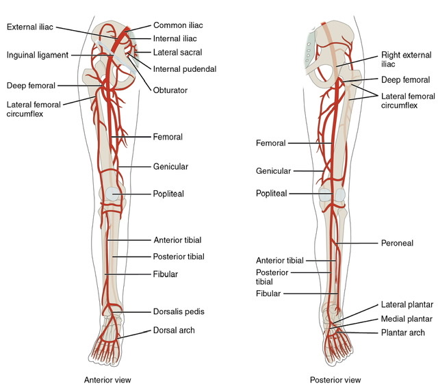
ARTERIAL SUPPLY OF LOWER LIMB
Q.1 What are the branches of the femoral artery?
The superficial branches include:
– Superficial circumflex iliac artery
– Superficial epigastric artery
– Superficial external pudendal artery
The deep branches include:
– Deep external pudendal artery
– Profunda femoris artery
– Descending genicular artery
Q.2 What is the extent of the femoral artery?
It begins at mid inguinal point and ends at the medial side of middle and lower one-third of the thigh by passing through an aperture in adductor magnus muscle to reach back of thigh and become popliteal artery.
Q.3 Name the branches of profunda femoris.
Q.4 Name the arteries forming the cruciate anastomosis.
Q.5 Name the arteries forming the trochanteric anastomosis.
Q.6 How the circulation is maintained in case of blockage of femoral artery?
In blockage in the proximal part, circulation is maintained through cruciate and trochanteric anastomosis. When the blockage is in lower thigh then circulation is maintained through perforating branches of the profunda femoris artery and its anastomoses with branches of the popliteal artery.
Q.7 Name the branches of the popliteal artery.
Q.8 What are the relations of the anterior tibial artery in anterior compartment of the leg?
- In upper 1/3, lies between tibialis anterior and extensor digitorum longus.
- In the middle 1/3, lies between tibialis anterior and extensor hallucis longus.
- In lower 1/3, lies between extensor hallucis longus and extensor digitorum longus.
Q.9 What are the branches of anterior tibial artery?
Q.10 How dorsalis pedis artery is formed?
It is the continuation of the anterior tibial artery in front of the ankle between the two malleoli.
Q.11 Name the branches of dorsalis pedis artery.
Q.12 Where the pulsations of dorsalis pedis artery are felt?
Between the tendon of extensor hallucis longus and first tendon of extensor digitorum longus on dorsum of foot about 5 cm distal to medial and lateral malleoli, over intermediate cuneiform bone.
Q.13 Name the branches of the posterior tibial artery.
Q.14 To which bone peroneal artery gives a nutrient artery?
Fibula
Q.15 Which artery forms the plantar arch?
Lateral plantar artery
Q.16 How the lateral plantar artery terminates?
It ends by joining the termination of dorsalis pedis artery in the interval between the bases of the first and second metatarsal bone.
VENOUS DRAINAGE
Q.1 What are the different factors that facilitate the return of venous blood to the heart?
– Veins of the lower limbs are larger than veins of other parts of the body. They also have a greater number of valves, which prevent the backflow of blood.
– Muscular contraction compresses the deep veins and drives the blood upwards.
– Muscular compression of veins is made more effective by tight deep fascia.
– The valves which maintain a unidirectional flow.
– Negative intrathoracic pressure, which pulls the column of blood up, and it is made more negative during inspiration.
– Vis-a-tergo (compulsion from behind) produced by arterial pressure and overflow from the capillary bed.
Q.2 What are the main superficial veins of the lower limb?
Q.3 What is ‘calf pump’ or ‘peripheral heart’?
In an upright position, venous return from lower limb depends largely on the contraction of calf muscles, these are known as calf pumps,& the soleus is called “peripheral heart” for same reason.
Q.4 What are varicose veins?
If the valves in veins become incompetent, the pressure during muscular contraction is transmitted from deep veins to the superficial veins and hence, leakage of blood. This causes dilatation of the superficial veins, known as varicose veins. Later on, gradual degeneration occurs, leading to “varicose ulcers”.
Q.5 What is the clinical importance of sural sinuses?
Sural sinuses are the common site for thrombosis and commonly leads to pulmonary embolism due to the detachment of thrombus.
LYMPHATIC DRAINAGE OF LOWER LIMB
Q.1 What is the lymphatic drainage of various inguinal lymph nodes?
Also read: Anatomy Question Collection
Also read: Anatomy Questions & Answers
Also read: Anatomy notes
Comments (0)