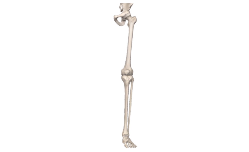
Joints of Lower Limb (Viva)
Hip Joint
Q.1 What is the type of hip joint?
Hip joint is a ball and socket type of synovial joint.
Q.2 What are the factors which increase the stability of the hip joint?
The stability of the hip joint is increased by the following factors:
- Depth of acetabulum with a narrow mouth, made by acetabular labrum.
- Tension and strength of ligaments.
- Strength of the surrounding muscles.
- Length and obliquity of the neck of femur.
The wide range of mobility depends upon the neck of the femur which is narrower than the equatorial diameter of the head.
Q.3 What is the attachment of the ligament of the head of the femur?
It is attached laterally to fovea on head of femur and medially to two ends of acetabular notch and to transverse ligament.
Q.4 What are the ligaments strengthening the capsule of the hip joint?
- Iliofemoral ligament: Strongest, Y-shaped ligament.
- Pubofemoral ligament
- Ischiofemoral ligament.
Q.5 What are the relations of the hip joint?
The relations of the hip joint are:
- Anteriorly: Lateral fibers are pectineus, iliopsoas, straight head of rectus femoris.
- Posteriorly: Quadratus femoris covering obturator externus and ascending branch of medial circumflex femoral artery, the piriformis, obturator internus with two gemelli separate the sciatic nerve from the nerve to quadratus femoris.
- Superior: Reflected head of rectus femoris covered by gluteus minimus.
- Inferior: Lateral fibers of pectineus and obturator externus.
Q.6 What is the blood supply to the hip joint?
The hip joint is supplied by the medial circumflex femoral and the lateral circumflex femoral vessels. There also may be a contribution by the acetabular branch of the femoral artery.
Q.7 What is the axis of different movements of the hip joint?
- For rotation, vertical axis passing through the center of the head of the femur and its lateral condyle.
- Extension and flexion, occur around a transverse axis.
- Adduction and abduction, occur around an anteroposterior axis.
Q.8 What is the range of movements at the hip joint?
Flexion is limited by contact of the thigh with the anterior abdominal wall.
Adduction is limited by contact with the opposite limb.
Range of other movements:
Lateral rotation 60°,
Medial rotation 25°,
Abduction 50° and extension 15°.
Q.9 What are the nerves supplying the hip joint?
The hip joint is supplied by:
- Femoral nerve, through nerve to rectus femoris,
- Anterior division of obturator nerve,
- Accessory obturator nerve,
- Nerve to quadratus femoris and
- Superior gluteal nerve.
Q.10 What are the different muscles producing extension of the hip joint?
Gluteus maximus and hamstrings.
Q.11 Which muscles produce abduction of the hip joint?
Chief muscles: Gluteus medius and minimus.
Accessory muscles: Tensor fasciae latae and sartorius.
Q.12 What is Trendelenburg test?
This test is employed for testing the stability of the hip joint.
A positive test indicates a defect in osseomuscular stability especially abductors of the hip joint and the patient has a “lurching” gait. If the patient is asked to stand on one leg. If the abductors of the thigh are paralyzed on that side, they will be unable to sustain the pelvis against the bodyweight and pelvis tilts downwards on an unsupported side.
Q.13 Name the adductors of the hip joint.
- Adductor longus
- Adductor brevis
- Adductor Magnus
- Gracilis
- Pectineus.
Q.14 Name the medial rotators of the hip joint.
Gluteus medius and minimus:
- Tensor fasciae latae
- Adductor longus, brevis, and Magnus.
Q.15 Name the flexors of the hip joint.
Mainly: Psoas major, iliacus, rectus femoris
Accessory muscles: Adductors are also flexors of the hip joint.
Q.16 What is the cause of Weaver’s bottom?
Inflammation of the bursa over the ischial tuberosity.
Q.17 In which injury of the hip joint sciatic nerve is likely to be damaged?
It is likely to be injured in the posterior dislocation of the hip joint associated with fracture of the posterior lip of the acetabulum, to which the nerve is closely related
KNEE JOINT
Q.1 What is the function of anterior and posterior cruciate ligament?
- Anterior cruciate ligament: Prevents hyperextension of the knee joint.
- Posterior cruciate ligament: Prevents hyperflexion of the knee joint.
Q.2 What is compartment syndrome?
It is an increase in fluid pressure (> 30 mm) within an osseofascial compartment and leads to muscle and nerve damage. Usually occur in the anterior compartment of the thigh as a result of crush injury can also occur in the anterior compartment of the leg due to fracture of the tibia.
Q.3 What is Legg-Perthes disease?
It is characterized by idiopathic avascular necrosis of the head of the femur. Caucasian boys are more commonly affected and it is usually characterised by unilateral hip pain external rotation (slight) and a limp.
Q.4 What type of joint is the knee joint?
Compound synovial joint, having
- Condylar synovial joint: Between the condyles of femur and tibia.
- Saddle synovial joint: Between femur and patella
Q.5 What are the articular surfaces in the knee joint?
Condyles of femur:
- Condyles of tibia and
- Patella
Q.6 What are the bony landmarks in the region of the knee?
- Adductor tubercle: is felt just above the medial condyle of the femur.
- Head of the fibula: is felt at the posterolateral aspect of the knee. Lies 1.5 cm below the level of the knee joint.
- Tibial tubercle: is felt in front of the knee in the upper part of the tibia.
- Tibial condyles: are felt on each side of the lower part of the ligamentum patellae.
Q.7 Name the ligaments of the knee joint.
- Fibrous capsule
- Ligamentum patellae
- Collateral ligaments: Tibial and fibular
- Popliteal ligaments: Oblique and arcuate
- Cruciate ligaments: Anterior and posterior
- Meniscus: Medial and lateral
- Transverse ligament
Q.8 What is coronary ligament?
It is the part of the fibrous capsule lying between the menisci and tibia.
Q.9 What are the openings in the fibrous capsule of the knee joint?
- For suprapatellar bursa and
- For the exit of tendon of popliteus with its synovial bursa.
Q.10 What are the attachments of arcuate popliteal ligament?
It passes from the head of the fibula to the posterior margin of the intercondylar area of the tibia.
Q.11 What are the attachments of oblique popliteal ligament?
It arises as an expansion from the tendon of semi-membranous. It blends with the posterior surface of the fibrous capsule. It is attached to the intercondylar line and lateral condyle of the femur and posterior aspect of the medial condyle of the tibia.
Q.12 Name the structures piercing oblique popliteal ligament.
- Posterior division of obturator nerve and
- Middle genicular nerve and vessels.
Q.13 What are menisci and what are their functions?
These are two fibrocartilaginous structures, semilunar in shape, which make the tibial articular surface deeper and divide the joint cavity partially into the upper and lower compartment.
Functions:
- They act as shock absorbers.
- They make the articular surfaces more congruent. They can adapt to varying curvatures of different parts of femoral condyles.
Q.14 What is the arterial supply of the knee joint?
- Genicular branches of popliteal artery,
- Descending genicular branch of femoral artery,
- Descending branch of the lateral circumflex femoral artery.
- Recurrent branches of the anterior tibial artery and
- Circumflex fibular branch of posterior tibial artery.
Q.15 Name the arteries forming the anastomosis around the knee joint.
Medially:
- Descending genicular
- Superior medial genicular
- Inferior medial genicular
Laterally:
- Descending branch of lateral circumflex femoral
- Superior lateral genicular
- Inferior lateral genicular
- Anterior lateral recurrent
- Posterior lateral recurrent
- Circumflex fibular
Q.16 What is the nerve supply of the knee joint?
- Femoral nerve,
- Genicular branches of tibial and common peroneal nerves and
- Posterior division of obturator nerve.
Q.17 What are the movements possible at the knee joint?
- Flexion
- Extension
- Medial and lateral rotation.
Q.18 What is ‘conjunct’ and ‘adjunct’ rotation?
- Conjunct rotation: Rotation of knee joint combined with flexion and extension.
- Adjunct rotation: Rotation of knee joint occurring independently in a partially flexed knee.
Q.19 What are the changes in the axis of movement of the knee joint with flexion and extension?
The flexion and extension of the knee joint takes place on a transverse axis which shifts along with the movements. Because of the spiral profiles of the femoral condyles, the axis shifts upwards and forwards during extension and backward and downwards during flexion.
Q.20 What are the locking and unlocking movements of the knee joint?
In full extension from the position of flexion, the last 30° of extension is accompanied by medial rotation of the femur on the tibia or lateral rotation of the tibia on the femur depending on whether the tibia or the femur is fixed. This is conjunct rotation and occurs passively as a part of the extension movement, is described as ‘locking’ of the knee joint. From the position of full extension, the beginning of flexion is accompanied by lateral rotation of the femur or medial rotation of the tibia depending on whether the tibia or the femur is fixed. This rotation is called ‘unlocking’ of the knee joint. The contraction of popliteus is responsible for this unlocking movement.
Q.21 Name the intra-articular structures of the knee joint.
- Cruciate ligaments: Anterior and posterior
- Menisci: Medial and lateral
- Infrapatellar pad of fat
- Synovial membrane
- Origin of popliteus
Q.22 Name the bursa around the knee joint?
Anteriorly:
- Subcutaneous prepatellar bursa
- Subcutaneous infrapatellar bursa
- Deep infrapatellar bursa and
- Suprapatellar bursa.
Medially:
- Bursa deep to the medial head of the gastrocnemius.
- Bursa deep to tibial collateral ligament.
- Semimembranosus bursa
- Anserine bursa and
- Occasionally, bursa between tendons of semitendinosus and semimembranosus.
Laterally:
- Bursa deep to the lateral head of the gastrocnemius
- Bursa between fibular collateral ligament and tendon of popliteus
- Bursa between fibular collateral ligament and biceps femoris and
- Bursa between tendon of popliteus and lateral condyle of tibia.
Q.23 Name the bursa communicating with the knee joint.
- Suprapatellar bursa
- Popliteal bursa
- Bursa deep to the medial head of the gastrocnemius
Q.24 What is Anserine bursa?
It is bursa with several diverticula which separate the tendons of sartorius, gracilis, and semitendinosus from the bony surface of the tibia.
Q.25 Name the different muscles producing movements of the knee joint.
| Movements | Principal muscles | Accessory muscles |
| Flexion | Semitendinosus, Biceps femoris, Semimembranosus |
Popliteus, Sartorius, Gastrocnemius, Gracilis |
| Extension | Quadriceps femoris | Tensor fasciae lata |
| Medial rotation | Semimembranosus Semitendinosus |
Sartorius, Gracilis |
| Lateral rotation | Biceps femoris |
Q.26 Name ligaments which become taut in full extension and flexion of the knee joint.
- In full extension:
– Anterior cruciate ligament.
– Tibial and fibular collateral ligament.
– Oblique popliteal ligament.
- In full flexion:
– Posterior cruciate ligament.
Q.27 What could cause a tear of the menisci (semilunar cartilages) of the knee joint?
The menisci are usually torn by a twisting force with knee flexed. When the flexed knee is forcibly abducted and externally rotated, the medial meniscus is trapped between the medial condyles of the femur and tibia and is torn.
A severe adduction and internal rotation of the flexed knee may result in a tear of the lateral meniscus. But this injury is less common.
Q.28 Why the tears of medial meniscus are more frequent than that of lateral meniscus?
Because the medial meniscus is more firmly attached to the upper surface of the tibia, capsule, and the tibial collateral ligament and therefore, is less able to adapt itself to sudden changes of position. The lateral meniscus, on the other hand, is drawn backward and downwards on the groove on the posterior aspect of the lateral tibial condyle by the medial fibers of popliteus. This prevents the lateral meniscus from being impacted between the articular surfaces of the femur and the tibia during movements of the knee joint.
Q.29 Why in tear of medial meniscus there is locking of the knee before it is fully extended?
Because the torn segment of the cartilage is displaced and lodges between the femoral and tibial condyles and prevents full extension of the knee.
Q.30 Why the pain of the hip joint is referred to as the knee?
Because of the common nerve supply of the two joints.
TIBIOFIBULAR JOINTS
Q.1 What type of joints are tibiofibular joints?
- Superior tibiofibular joint: Plane synovial joint.
- Lower tibiofibular joint: Syndesmosis type of fibrous joint.
Q.2 Name the structures passing through the interosseous membrane of the tibiofibular joint.
- Anterior tibial vessels
- Perforating branch of the peroneal artery.
ANKLE JOINT
Q.1 What type of joint is the ankle joint?
Hinge variety of synovial joint
Q.2 What are the articular surfaces of the ankle joint?
- From above:
– Lower end of tibia with medial malleolus
– Lateral malleolus and
– Inferior transverse tibiofibular ligament.
- From below:
Body of talus
Q.3 Name the ligaments of the ankle joint.
- Fibrous capsule
- Lateral ligament: Consists of
– Anterior talofibular ligament,
– Posterior talofibular ligament and
– Calcaneofibular ligament.
- Medial (Deltoid) ligament: It has
- Superficial part: Consists of
– Anterior fibers (Tibionavicular)
– Middle fibers (Tibiocalcanean) and
– Posterior fibers (Posterior tibiotalar). - Deep part (Anterior tibiotalar)
Q.4 Name the tendons crossing the deltoid ligament.
- Tibialis posterior and
- Flexor digitorum longus.
Q.5 Name the structures related to ankle joint.
Anteriorly:
From medial to lateral side:
- Tibialis anterior.
- Extensor hallucis longus,
- Anterior tibial vessels,
- Deep peroneal nerve,
- Extensor digitorum longus and
- Peroneus tertius.
Posteriorly:
From medial to lateral side
- Tibialis posterior,
- Flexor digitorum longus,
- Posterior tibial vessels,
- Tibial nerve,
- Flexor hallucis longus
- Peroneus brevis and
- Peroneus longus
Q.6 What are the movements produced at the ankle joint?
- Dorsiflexon
- Plantar flexion
- Accessory movements:
With plantar flexion, a slight amount of side to side gliding, abduction and adduction are permitted.
Q.7 What is the axis of movements of the ankle joint?
It is represented by a transverse line drawn across the front of the ankle about 1.25 cm above the tip of the medial malleolus.
Q.8 What is the close-pack position of the ankle joint?
Dorsiflexion is the close-pack position of the joint in which the wider front part of the talus articulates with the mortise formed by the malleoli and lower end of the tibia. In this position, there is the maximal congruence of the joint surface and tension of the ligaments.
Q.9 Name the muscles producing movements at ankle joint.
Dorsiflexion:
- Main muscle: Tibialis anterior
- Accessory muscles:
– Externsor digitorum longus,
– Extensor hallucis longus and
– Peroneus tertius.
Plantar flexion:
- Main muscles:
– Gastrocnemius and
– Soleus
- Accessory muscles:
– Flexor digitorum longus,
– Flexor hallucis longus,
– Tibialis posterior and
– Plantaris
Q.10 What is the most frequent fracture at the ankle joint?
Pott’s fracture, usually produced by an abduction external rotation injury
JOINTS OF FOOT
Q.1 What do you understand by inversion and eversion of foot?
Inversion:
Movement in which the medial border of the foot is elevated and sole faces medially and inwards.
Eversion:
Movement in which the lateral border of the foot is elevated and sole faces laterally and outwards.
Q.2 Name the joints at which inversion and eversion takes place.
- Subtalar (Talocalcaneal) joint and
- Talocalcaneonavicular joint
Q.3 What is the axis of the inversion and eversion?
Oblique axis which runs forwards, upwards and medially. It passes between the back of calcaneum, sinus tarsi, and the superomedial aspect of the neck of the talus.
Q.4 Name the evertors of foot.
- Mainly by, peroneus brevis and longus.
- Also by, peroneus tertius.
Q.5 Name the invertors of foot.
Principal muscles:
- Tibialis anterior and
- Tibialis posterior.
Accessory muscles:
- Flexor hallucis longus and
- Flexor digitorum longus
Q.6 Why are the movements of inversion and eversion required in man?
Inversion and eversion of the foot are essential for walking on rough, uneven or sloping surfaces.
Also read: Anatomy Question Collection
Also read: Anatomy Questions & Answers
Also read: Anatomy notes

Comments (0)