Femoral Triangle (Viva)
6 years ago 5312
FEMORAL TRIANGLE
Q.1 Why femoral triangle is known as Scarpa’s triangle?
Because it was first described by Antonio Scarpa (1747-1832) in Italy.
Q.2 What are the boundaries of the femoral triangle?
It is bounded by
Q.3 What are the contents of the femoral triangle?
Q.4 What is femoral sheath?
It is a funnel-shaped fascial sleeve enclosing the upper 1½ inches of the femoral vessels.
Q.5 How is femoral sheath formed?
It is formed by the downward extension of the abdominal fasciae. The anterior wall is formed by fascia transversalis and posterior wall by fascia iliaca.
Q.6 What are the relations of the femoral sheath?
Anterior:
Posterior:
Lateral:
Medial:
Q.7 What are the parts of the femoral sheath?
The cavity within the femoral sheath is divisible in three parts.
Lateral part contains the femoral artery and a femoral branch of the genitofemoral nerve.
Middle part contains the femoral vein and the medial part is called the femoral canal.
Q.8 What is the femoral canal?
It is the medial compartment of the femoral sheath. It is conical and ½ inch wide at the base and ½ inch long.
Q.9 What is the femoral ring?
The base or upper end of the femoral canal is called the femoral ring. The femoral ring is filled by condensed extraperitoneal tissue, the femoral septum, containing a lymph node and covered by parietal peritoneum.
Q.10 What are the boundaries of the femoral ring?
Q.11 What are the contents of the femoral canal?
Q.12 What are the functions of the femoral canal?
Q.13 What structure is drained by the lymph node of the femoral canal?
Glans penis in the male and clitoris in females.
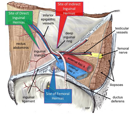
Q.14 What is the clinical importance of the femoral canal?
The femoral canal is a potential point of weakness in the lower abdominal wall through which a viscus (intestines or urinary bladder) may protrude and give rise to a femoral hernia.
Q.15 Why is a femoral hernia commoner in females?
Because the femoral canal is larger in the females due to the greater width of the pelvis and smaller size of the femoral vessels. In the females, there is a rise in intraabdominal pressure due to pregnancy predisposing to femoral hernia.
Q.16 Why is strangulation more common in femoral hernia?
Because the neck of the femoral canal is narrow.
Q.17 What is the risk of enlarging the opening of the femoral canal in releasing the strangulation of a femoral hernia?
In order to enlarge the opening of the femoral canal the sharp lateral edge of the lacunar (Gimbernat’s) ligament may require an incision. An abnormal obturator artery may occasionally be present, which passes behind the lacunar ligament and is then in danger of being cut.
Q.18 What are the coverings of femoral hernia?
From within outwards:
Also read: Anatomy Question Collection
Also read: Anatomy Questions & Answers
Also read: Anatomy notes
Comments (0)