Bones of Foot & Arches of Foot (Viva)
6 years ago 3528
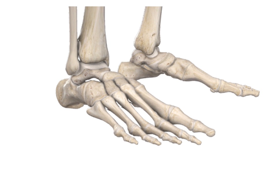
BONES OF FOOT
Q.1 Name the tarsal bone of the foot.
Navicular is interposed between the two rows.
Q.2 Name the structures attached to the medial tubercle of the calcaneum.
Medially:
Q.3 Name the structures attached to the lateral tubercle of the calcaneum.
Origin of abductor digiti minimi.
Q.4 Name the tendons related to peroneal trochlea of calcaneum.
Above: Tendon of peroneus brevis
Below: Tendon of peroneus longus.
Q.5 What are the structures attached to sustentaculum tali?
To its medial margin are attached
Q.6 What is the structure attached to the tuberosity of navicular bone?
Insertion for tibialis posterior
Q.7 Name the structures related to the plantar groove of cuboid.
Q.8 At what time the ossification center for cuboid appears?
Just before or after birth.
Q.9 What are the differences between metacarpal and metatarsal?
| Regions | Metacarpal | Metatarsal |
| Head and shaft: | Prismoid | Flattened from side to side |
| Shaft: | Uniform thickness | Tapers distally |
| Dorsal surface of shaft: | Elongated, flat triangular area | Uniformly convex |
| Base: | Irregular | Cuts sharply and obliquely |
Q.10 What are the “accessory bones”?
These are separate small pieces of bone which have not fused with the main bone e.g.,
Q.11 What is ‘bunion’?
It is inflamed adventitial bursa over the head of the first metatarsal bone.
ARCHES OF FOOT
Q.1 Classify the arches of the foot.
Q.2 How the arches of the foot are maintained?
Q.3 What are the functions of arches of foot?
Q.4 How the medial longitudinal arch is formed?
By calcaneum, talus, three cuneiforms, and three medial metatarsals.
The summit of the arch is formed by talus.
Q.5 How the lateral longitudinal arch is formed?
By the calcaneum, cuboid and lateral two metatarsals.
Q.6 How the transverse arch is formed?
By the bases of the five metatarsals and the adjacent cuboid and cuneiforms of both feet.
Q.7 What are the attachments of spring ligament?
It passes from anterior magin of sustentaculum tali of calcaneus to plantar surface of navicular bone.
Q.8 What are the attachments of long plantar ligament?
It is attached posteriorly to the plantar surface of the calcaneus in front of lateral and medial tubercles and anteriorly to the plantar surface of cuboid distal to groove for peroneus longus.
Q.9 Which structures maintain the medial longitudinal arch?
The bony configuration does not contribute to the maintenance of this arch.
– The medial part of the plantar aponeurosis acts as a tie beam.
– The plantar calcaneonavicular (‘spring’) ligament supports the head of the talus and forms intersegmental ties (connect adjacent bones).
– Medial half of the flexor digitorum brevis and abductor hallucis act as tie beams (connect ends of arch)
– Tibialis anterior, tibialis posterior, and flexor hallucis longus act by forming sling and suspend the arch.
Q.10 How the lateral longitudinal arch of the food is maintained?
– The short plantar ligament, long plantar ligament and dorsal ligaments form intersegmental ties.
– Lateral part of the plantar aponeurosis acts as a tie beam.
– The peroneus longus and peroneus brevis muscles form the slings.
– Lateral half of the flexor digitorum brevis and abductor digiti minimi act as tie beam.
Q.11 How the transverse arch of the foot is maintained?
Tarsal and metatarsal bones contribute to maintaining the concavity of the arch.
– Ligaments that bind together the cuneiforms and the bases of the metatarsals form intersegmental ties.
– Superficial and deep transverse metatarsal ligaments act as tie beams.
– The peroneus longus and tibialis posterior form slings.
– Abductor hallucis acts as a tie beam.
Q.12 What are the deformities of the foot resulting from defects of the longitudinal arches of the foot?
Q.13 What is the ‘talipes deformity’ of the foot?
In talipes, the foot no longer lies in the plantigrade position. The person walks either on the heels or on the toes. When he walks on the heel the condition is known as talipes calcaneus while walking on the toes is known as talipes equinus. In both these conditions, the foot may be inverted (varus) or everted (valgus).
Q.14 What is Hallus valgus?
In hallux valgus, there is a lateral deviation of the great toe at the metatarsophalangeal joint. More common in women than men.
Q.15 What is ‘Hammer toe’?
The affected toe is hyperextended at the metatarsophalangeal and distal interphalangeal joint and flexed at the proximal interphalangeal joint.
Also read: Anatomy Question Collection
Also read: Anatomy Questions & Answers
Also read: Anatomy notes
Comments (0)