Tibia, Fibula & Patella (Viva)
6 years ago 3684
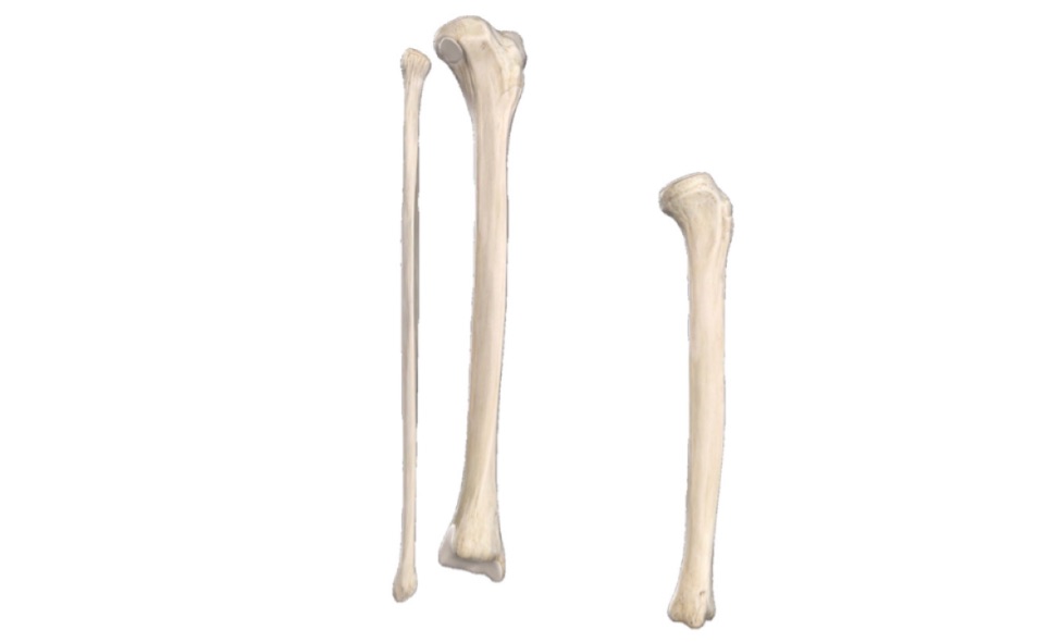
TIBIA AND FIBULA
Q.1 Name the structures attached to the intercondylar area of the tibia.
From before backward, it provides attachment to:
Q.2 What are the structures related to the anterior surface of the lower end of the tibia?
From medial to lateral side it is related to tibialis anterior, extensor hallucis longus, anterior tibial vessels, deep peroneal nerve, and extensor digitorum longus.
Q.3 What are the structures related to the posterior surface of the lower end of the tibia?
From medial to lateral side it is related to tibialis posterior, flexor digitorum longus, posterior tibial artery, tibial nerve, and flexor hallucis longus.
Q.4 What is the arterial supply of tibia?
The nutrient artery to the tibia is a branch of the posterior tibial artery. It is the largest nutrient artery in the body.
Q.5 Although the tibia is one of the commonest sites of acute osteomyelitis but the knee joint is not involved. Explain?
The knee joint is not involved because the capsule is attached near articular margins of the tibia, proximal to the epiphyseal line.
Q.6 The fracture of tibia is slow healing. Why?
The tibia is commonly fractured at the junction of upper 2/3 and lower 1/3 of its shaft, where it is most slender and this site is poorly supplied by blood vessels.
Q.7 How will you determine the side to which the fibula belongs?
The head is slightly expanded in all directions and lateral malleolus is expanded anteroposteriorly and is flattened from side to side. The medial side of lower end bears a triangular articular facet anteriorly and malleolar fossa posteriorly.
Q.8 Which structure lies between two heads of origin of peroneus longus?
Common peroneal nerve.
Q.9 Name the structures attached to malleolar fossa.
Malleolar fossa provides attachment to the posterior talofibular and posterior tibiofibular ligament.
Q.10 Fibula violates the general rule of ossification. Explain.
Normally in a long bone, the growing end of a long bone ossifies first and unites with the shaft last while the non-growing end ossifies last and fuses with the shaft first. But in fibula, the ossification center for nongrowing end, i.e. lower end appears first but does not fuse last.
This occurs because:
Q.11 What are the functions of fibula?
PATELLA
Q.1 What is the function of the patella?
The patella improves the leverage of the quadriceps femoris by increasing the angulation of the line of pull on the leg.
Q.2 How the stability of the patella is increased?
Due to outward angulation between long axes of thigh and leg the patella has a tendency to dislocate outwards.
This is prevented by:
Q.3 What are the different sesamoid bones present in the lower limb?
The following sesamoid bones are present in the lower limb:
Q.4 What is ‘Febella’?
It is a small, rounded sesamoid bone present in the lateral head of gastrocnemius. It articulates with the posterior surface of the lateral condyle of the femur.
Also read: Anatomy Question Collection
Also read: Anatomy Questions & Answers
Also read: Anatomy notes
Comments (0)