Femour & Thigh (Viva)
6 years ago 3504
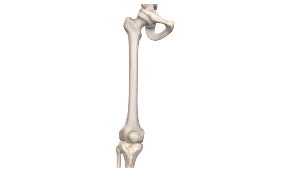
Femur
Q.1 What is the normal anatomical position of the femur in the body?
The head of the femur is directed medially, upwards and slightly forwards and the shaft is obliquely downwards and medially so that the two condyles at lower surface lie in the same the horizontal plane.
Q.2 What is the arterial supply of the head of the femur?
The medial part near the fovea, supplied by medial epiphyseal arteries derived from ascending branch of the medial circumflex femoral artery and posterior division of obturator artery. The lateral part of the head is supplied by lateral epiphyseal arteries derived from the lateral circumflex femoral artery.
Q.3 What is the nutrient artery of the femur?
It is derived from the second perforating artery.
Q.4 What is angle of anteversion?
The angle of anteversion (angle of femoral torsion) is the angle between the transverse axes of upper and lower ends of the femur. It is about 15 degrees.
Q.5 Name the structures attached to the intertrochanteric line of femur.
The following structures are attached to the intertrochanteric line:
Q.6 Which muscle is inserted into trochanteric fossa?
Obturator externus.
Q.7 Which muscle is inserted in gluteal tuberosity?
Deep fibers of gluteus maximus.
Q.8 What is the origin of popliteus muscle?
From anterior part of groove on lateral aspect of lateral condyle of femur.
Q.9 What is the importance of the ossification center for the lower end of the femur?
The ossification center for the lower end of the femur appears at end of 9th month of intrauterine life (the day of birth). It is of medicolegal importance in cases of newly born child found dead to decide whether it was viable or not.
Q.10 What is characteristic of the primary ossification center of femur?
It is the second long bone in the body to start ossifying.
Q.11 Why the fractures of the neck of femur, lead to the necrosis of the head?
Because it will interrupt the blood supply to the head which is derived from:
Q.12 Why the intracapsular fracture of the neck of the femur are more dangerous than extracapsular fracture?
The intracapsular fracture interrupts the blood supply, to the femoral head resulting in necrosis whereas in the extracapsular fracture, the blood supply to the head remains unaffected and so there is no danger of avascular necrosis.
Q.13 What is Coxa vara?
In this condition, the angle between the femoral neck and shaft is decreased i.e., less than 160°. This results from adduction fractures.
Q.14 What is Coxa valga?
Increase in the angle between femoral neck and shaft due to abduction fractures.
Q.15 At which level fracture of shaft of femur is dangerous?
Fracture of the lower end of the femur is dangerous because the proximal edge of the distal fragment is tilted backward by the gastrocnemius, which tears the popliteal artery which lies directly behind it.
THIGH
Q.1 What is the mid-inguinal point and what is its importance?
Midinguinal point is a point midway between anterior superior iliac spine and the pubic symphysis. It is an important landmark. The femoral artery and head of the femur lie beneath the mid inguinal point.
Q.2 What is Holden’s line and what is its importance?
The deep layer of superficial fascia is firmly attached to the deep fascia of the thigh along a horizontal line a little lateral to the pubic tubercle and extends for about 8 cm laterally. This line of firm attachment is called Holden’s line. Clinical importance: The extravasation of urine between these two layers cannot extend into the thigh because of the firm attachment.
Q.3 How is patellar plexus formed?
It is a plexus of nerves in front of the patella and upper end of tibia.
It is formed by
Q.4 What is Housemaid’s knee?
Chronic enlargement of prepatellar bursa is known as Housemaid’s knee because it commonly occurs in housemaid’s who have to kneel regularly for sweeping the floor.
Q.5 What is Miner’s beat knee?
It is acute suppurative prepatellar bursitis in miners.
Q.6 What is Clergyman’s knee?
It is an enlargement of the subcutaneous infrapatellar bursa in the clergyman.
Q.7 What is iliotibial tract and what is its function?
The thickening of fascia lata on the lateral side of the thigh is called the iliotibial tract.
Functions:
Q.8 What are the modifications of the deep fascia of the thigh?
Also read: Anatomy Question Collection
Also read: Anatomy Questions & Answers
Also read: Anatomy notes
Comments (0)