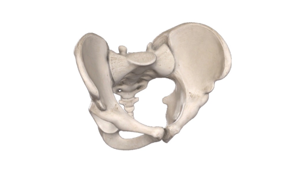
Hip Bone & Gluteal Region (Viva)
HIP BONE
Q.1 What are the different parts of a hip bone?
The hip bone is made up of three parts, the ilium superiorly, ischium postero-inferiorly, and pubis antero-inferiorly. The three parts join to form a cup-shaped hollow articular surface, the acetabulum.
Q.2 How will you determine to which side the hip bone belongs?
In a hip bone, the acetabulum is directed laterally and the flat ilium forms the upper part of the bone, lying above the acetabulum. the obturator foramen lies below the acetabulum.
Q.3 What is the normal anatomical position of the hip bone in the body?
- Pubic tubercle and anterior superior iliac spine lie in the same vertical plane.
- The pelvic surface of the body of pubis is directed backward and upwards.
- The ischial spine and upper border of symphysis pubis lie in same horizontal plane and
- Symphysis pubis lies in the median plane.
Q.4 What is the level through which the highest point of the iliac crest passes (intercrestal plane)?
The intercrestal plane passes at the level of the interval between the spines of L3 and L4 vertebrae.
Q.5 What is the clinical importance of intercrestal plane?
In clinical practice, lumbar puncture is done between the L3 and L4 vertebrae.
Q.6 What are the structures attached to the anterior superior iliac spine?
It provides:
- Attachment to the lateral end of inguinal ligament and
- Origin of Sartorius.
Q.7 Name the structures attached to the iliac crest.
Anterior 2/3 of iliac crest has:
- Outer lip which provides
– Attachment of fascia lata,
– Origin of tensor fasciae lata,
– Insertion to external oblique muscle and
– Origin to latissimus dorsi just behind the highest point.
- Intermediate area provides origin to internal oblique muscle.
- Inner lip provides
– Origin to transversus abdominis,
– Attachment to fascia iliaca and fascia transversalis,
– Origin to quadratus lumborum in posterior 1/3 and
– Attachment to thoracolumbar fascia.
Posterior 1/3 segment of iliac crest has:
- Lateral slope: Origin of gluteus maximus.
- Medial slope: Origin of erector spinae.
- Medial margin: Interosseous and dorsal sacroiliac ligaments.
Q.8 Name the structures attached to the anterior inferior iliac spine.
Anterior inferior iliac spine gives:
- Origin to straight head of rectus femoris in superior half and
- Attachment to iliofemoral ligament in inferior half.
Q.9 Name the structures attached to the posterior border of the ilium.
It provides:
- Attachment to upper fibers of sacrotuberous ligament and
- Origin to fibers of piriformis.
Q.10 What are the structures attached to the gluteal surface of ilium?
- Gluteus medius arises between anterior and posterior gluteal lines.
- Gluteus minimus arises between anterior and inferior gluteal lines.
- Gluteus maximus (upper fibers) arise behind the posterior gluteal line.
- Below the inferior gluteal line reflected head of rectus femoris arises.
Q.11 Name the structures attached to the pubic tubercle.
- Medial end of the inguinal ligament.
- Ascending loops of cremaster muscle.
Q.12 Name the structures attached to the crest of the pubis.
- Lateral head of rectus abdominis (origin)
- Pyramidalis (origin).
Medial head of rectus abdominis arises from the anterior pubic ligament.
Q.13 What are the structures attached to the pectineal line?
The structures attached to the pectineal line are:
- Conjoint tendon and lacunar ligament at medial end.
- Pectineal ligament lateral to lacunar ligament.
- Origin of pectineus muscle and fascia covering it, from the whole length.
- Insertion of psoas minor.
Q.14 Name the structures attached to the ischial spine.
The structures attached to the ischial spine are:
- Sacrospinous ligament
- Origin of coccygeus and levator ani.
- Origin of superior gemellus
Q.15 What are the structures attached to ischial tuberosity?
From upper area of ischial tuberosity arise semimembranous superolaterally and semitendinosus and long head of biceps femoris superomedially.
From the lower lateral area Abductor Magnus arise.
Q.16 What are the nerves related to hip bone?
- Sciatic nerve related to the lower margin of the greater sciatic notch.
- Obturator nerve in the obturator canal.
- Nerve to obturator internus crosses the base of the ischial spine.
- Pudendal nerve crosses the base of the ischial spine.
- Nerve to quadratus femoris runs on ischium as it crosses the greater sciatic notch.
GLUTEAL REGION
Q.1 Name the structures passing through the greater sciatic foramen.
Piriformis
- Structures passing above piriformis
– Superior gluteal nerve
– Superior gluteal vessels
- Structures passing below piriformis
– Inferior gluteal vessels
– Internal pudendal vessels
– Inferior gluteal nerve
– Sciatic nerve
– Posterior cutaneous nerve of thigh
– Nerve to quadratus femoris
– Pudendal nerve
– Nerve to obturator internus.
Q.2 Name the structures passing through the lesser sciatic foramen.
- Tendon of obturator internus
- Internal pudendal vessels
- Pudendal nerve
- Nerve to obturator internus.
Q.3 Name the structures lying under cover of gluteus minimus.
- Reflected head of rectus femoris
- Capsule of hip joint.
Q.4 What are the structures lying under the cover of the gluteus medius?
- Superior gluteal nerve
- Deep branch of superior gluteal artery
- Gluteus minimus
- Trochanteric bursa of gluteus medius.
Q.5 Name the structures lying under the cover of gluteus maximus.
- Ligaments
– Sacrotuberous
– Sacrospinous and
– Ischiofemoral
- Bones and joints
– Ilium
– Ischium with ischial tuberosity
– Upper end of the femur with the greater trochanter
– Sacrum
– Coccyx
– Hip joint
– Sacroiliac joint.
- Bursae
– Trochanteric bursa of gluteus maximus
– Bursa over ischial tuberosity and
– Bursa between gluteus maximus and vastus lateralis.
- Muscles
– Gluteus medius
– Gluteus minimus
– Reflected head of rectus femoris
– Piriformis
– Obturator internus
– Superior and inferior Gemelli
– Quadratus femoris
– Obturator externus
– Origin of hamstrings
– Insertion of adductor Magnus.
- Vessels
– Superior gluteal vessels
– Inferior gluteal vessels
– Internal pudendal vessels
– Ascending branch of the medial circumflex femoral artery
– Trochanteric anastomosis
– Cruciate anastomosis
– First perforating artery.
- Nerves
– Superior gluteal (L4,5 S1)
– Inferior gluteal (L5, S1,2)
– Sciatic (L4,5 S1,2,3)
– Posterior cutaneous nerve of thigh (S1,2,3)
– Nerve to quadratus femoris (L4,5 S1)
– Pudendal nerve (S2,3,4)
– Nerve to obturator internus (L5, S1,2)
– Perforating cutaneous nerve (S2,3)
Q.6 What is Waddling gait?
Results from bilateral paralysis of gluteus medius and minimus so that the patient walks with swaying to clear the feet off the ground.
When unilateral then it is known as lurching gait.
Also read: Anatomy Question Collection
Also read: Anatomy Questions & Answers
Also read: Anatomy notes

Comments (0)