Bones of Hand & Space of Hand (Viva)
6 years ago 2890
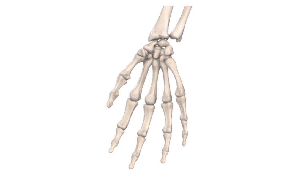
BONES OF HAND
Q.1 Name the carpal bones.
Proximal row:
Contains from lateral to medial side
Distal row:
Contains from lateral to medial side
Q.2 Name the structures attached to the tubercle of the scaphoid.
Q.3 Name the structures attached to pisiform.
Q.4 Name the site of the insertion of various muscles of the thumb.
Q.5 Name the muscles attached to the middle phalanx.
Q.6 Name the structures attached to the base of the proximal phalanx.
Insertion of lumbricals and interossei.
Insertion of
– On the lateral side: Abductor pollicis brevis and flexor pollicis brevis.
– On medial side: Abductor pollicis and first palmar interosseous.
– On dorsal surface: Extensor pollicis brevis.
On medial side:
Insertion of abductor digiti minimi and flexor digiti minimi.
Q.7 What are the sesamoid bones found in the upper limb?
Spaces of Hand
Q.1 Name the spaces of hand.
– Superficial pulp spaces of the fingers
– Synovial tendon sheats of 2nd, 3rd and 4th fingers
– Ulnar bursa
– Radial bursa
– Midpalmar space
– Thenar space.
– Dorsal subaponeurotic space
– Dorsal subcutaneous space.
Q.2 What are the characteristic features of the pulp space of fingers?
Q.3 Why the infections of pulp space of fingers are painful?
Because it cannot expand due to fibrous processes attaching skin to the periosteum and thus little swelling causes much increase in tension.
Q.4 Why the infections of pulp space of fingers cause necrosis of distal 4/5 of terminal phalanx?
Because distal 4/5 receives its blood supply from arteries which transverse fibrous processes and increase in tension due to infection causes their occlusion and proximal 1/5 escapes necrosis because it receives its blood supply by vessels which do not traverse fibrous processes.
Q.5 Why the infections of little finger and thumb are more dangerous?
Because the synovial sheath of the little finger is continuous with ulnar bursa and that of thumb with radial bursa so, the infections of these can spread of the forearm space of Parona.
Q.6 What is the position of dorsal spaces?
The subcutaneous space lies deep to the skin and sub-aponeurotic space deep to extensor tendons on the dorsal aspect of the hand.
Q.7 What is the `forearm space of Parona' and its clinical importance?
It is space between long flexor tendons and pronator quadratus. Proximally, the upward extent is limited by the origin of flexor digitorum superficialis and inferiorly, it extends up to the upper border of flexor retinaculum. The proximal parts of the flexor tendons synovial sheath protrude into it.
Clinical importance:
It may be infected by the extension of synovial sheath infections from ulnar or radial bursa, leading to hourglass swelling.
Q.8 What is the position and clinical importance of mid-palmar and thenar space?
These are potential spaces deep to palmar aponeurosis and flexor tendons.
Q.9 Why the collection of fluid is more on the dorsal surface of hand in infections of the palmar aspect of fingers?
Skin on dorsum of fingers and hand is loose, therefore fluid readily collects beneath it. But on the palm of the hand, there is little subcutaneous tissue and skin is adherent to the underlying palmar fascia.
Also read: Anatomy Question Collection
Also read: Anatomy Questions & Answers
Also read: Anatomy notes
Comments (0)