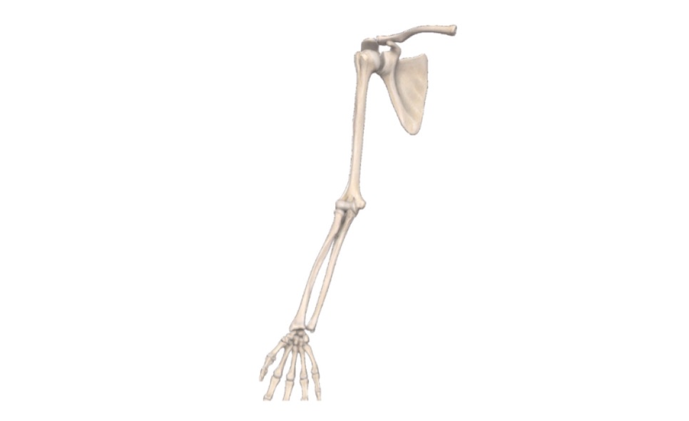
Joints of Upper Limb (VIva)
SHOULDER JOINT
Q.1 What type of joint shoulder joint is?
- Ball and socket variety of synovial joint
Q.2 What are the articular surfaces of the shoulder joint?
- The glenoid cavity of scapula and head of humerus.
Q.3 Name the ligaments of the shoulder joint?
- Capsular ligament.
- Coracohumeral ligament.
- Transverse humeral ligament.
- Glenoid labrum.
Q.4 What is the arterial supply of the shoulder joint?
- Anterior circumflex humeral artery.
- Posterior circumflex humeral artery.
- Suprascapular artery and
- Subscapular artery
Q.5 What are the movements possible at the shoulder joint? Name also the main muscle producing these movements?
- Flexion:
- Clavicular head of pectoralis major
- Anterior fibres of deltoid.
- Coracobrachialis
- Short head of biceps
- Extension:
- Sternocostal head to pectoralis major
- Posterior fibers of deltoid
- Latissimus dorsi
- Teres major.
- Adduction:
- Pectoralis major
- Latissimus dorsi
- Subscapularis
- Teres major.
- Abduction:
- Middle fibers of deltoid
- Supraspinatus
- Medial rotation:
- Pectoralis major
- Anterior fibres of deltoid
- Latissimus dorsi
- Teres major
- Subscapularis.
- Lateral rotation:
- Posterior fibers of deltoid
- Infraspinatus
- Teres minor.
- Circumduction: Combination of different movements.
Q.6 Name the bursa around the shoulder joint.
- Subacromial bursa
- Subscapularis bursa
- Infraspinatus bursa
- Bursa related to muscles around the shoulder joint, e.g. teres major, long head of triceps, coracobrachialis
Q.7 a) Why shoulder joint is a weak joint?
b) How its stability is increased?
a)
- The glenoid cavity is shallow and small.
- The Head of humerus is larger than the glenoid cavity.
b)
- By musculotendinous cuff of the shoulder.
- Coracoacromial arch.
- Glenoidal labrum, which deepens the glenoid cavity and articular cartilage lining it.
- Long muscles of shoulder, e.g. deltoid, long head of triceps, latissimus dorsi and teres major.
Q.8 What is ‘Rotator cuff’ or ‘Musculotendinous cuff’?
- It is a fibrous sheath of tendons of short muscles of the shoulder which cover all except inferior aspects of the shoulder joint. The muscles are supraspinatus (superiorly) subscapularis (anteriorly), infraspinatus, and teres minor (posteriorly). The cuff gives strength to the capsule of the shoulder joint.
Q.9 Which tendon is most commonly injured in rotator cuff lesions?
- Supraspinatus
Q.10 Why the dislocation of the shoulder joint occurs inferiorly?
- Because the inferior aspect is unprotected by musculotendinous cuff.
Q.11 What is the clinical importance of shoulder tip pain?
- Irritation of the undersurface of the diaphragm from surrounding pathology causes referred pain in the shoulder because the phrenic nerve and supraclavicular nerves have similar root values (C3,4).
- Pain in the left shoulder tip due to irritation by splenic rupture.
- Pain in the right shoulder, due to subphrenic abscess.
- Acute pancreatitis and gas under the diaphragm due to perforation of peptic ulcer causes referred pain in the either of the shoulder tip.
SHOULDER GIRDLE
Q.1 What are the joints of the shoulder girdle?
- Sternoclavicular joint
- Acromioclavicular joint.
Q.2 What type of joints are joints of shoulder girdle?
- Sternoclavicular joint: Saddle variety of synovial joints.
- Acromioclavicular joint: Plane variety of synovial joint.
Q.3 What is the characteristic feature of the acromioclavicular joint?
- It is partially divided by an incomplete fibrocartilage articular disc, which is perforated in the center.
Q.4 Name the ligaments forming acromioclavicular joint?
- Coracoclavicular ligament: Main ligament
- Coracoacromial ligament
Q.5 What are the movements produced at the shoulder girdle?
- Elevation of scapula:
- By upper fibers of trapezius and
- Levator scapulae. For example, shrugging of shoulders.
- Depression of scapula: By
- Lower fibers of serratus anterior
- Pectoralis minor
- Levator scapulae and rhomboids also assist.
- Protraction of the scapula: By
- Serratus anterior and
- Pectoralis minor. For example, Punching movements.
- Retraction of scapula: By
- Rhomboids and
- Middle fibers of trapezius.
- Forward rotation of scapula around chest wall: In overhead abduction of shoulder by:
- Upper fibers of trapezius and
- Lower fibers of serratus anterior.
- Backward rotation of scapula: By
- Levator scapulae and
- Rhomboids
Q.6 What is the function of the shoulder girdle?
- It suspends the upper limb to the axial skeleton
Q.7 What type of joint elbow joint is?
Hinge variety of synovial joint
Q.8 What are the surfaces of the elbow joint?
From above:
Capitulum and trochlea of humerus
From below:
- Capitulum articulates with the upper surface of head of radius
- The trochlear notch of the ulna articulates with the trochlea of the humerus
Q.9 Name the ligaments of the elbow joint.
- Capsular ligament.
- Anterior ligament.
- Posterior ligament.
- Ulnar collateral ligament.
- Radial collateral ligament.
Q.10 What are the movements of the elbow joint? Name the muscles producing these movements.
Flexion: By
- Brachialis,
- Biceps and
- Brachioradialis.
Extension: By
- Triceps and
- Anconeus.
Q.11 How will you clinically test for dislocation of elbow joint?
- Normally, in a semi flexed position, olecranon and two humeral epicondyles form an equilateral triangle. In the dislocation of the elbow, this relationship is disturbed.
Q.12 What is ‘Tennis Elbow’?
- It is due to the partial tear of the common origin of the superficial extensor muscles of the forearm.
Q.13 What is ‘Golfer's Elbow’?
- It is due to a partial tear of the common origin of the superficial flexor muscles of the forearm.
Q.14 What is the student’s/miner’s elbow?
- Repeated pressure over the olecranon process leading to inflammation of olecranon bursa.
RADIOULNAR JOINTS
Q.1 What type of joint radioulnar joints are?
- Superior radioulnar joint: Pivot type of synovial joint.
- Inferior radioulnar joint: Pivot type of synovial joint.
- Middle radioulnar joint: Syndesmoses type of fibrous joint.
Q.2 What are the functions of the interosseous membrane of the middle radioulnar joint?
- Attachment to muscles,
- Transmits weight of hand from radius to ulna.
Q.3 What is pronation and supination?
- These are rotatory movements of the forearm with the hand around a vertical axis in a semi flexed position.
- In pronation, palm faces downwards
- In supination, palm faces upwards.
Q.4 What is the axis of pronation and supination?
- Vertical axis passing superiorly, through the centre of the head of radius and inferiorly, through the apex of the articular disc when ulna is fixed or through any fixed finger when ulna is free to move.
Q.5 Name the muscles producing pronation and supination.
- Pronation: Principal muscles: Pronator teres, Pronator quadratus. Accessory muscles: Flexor carpi radialis, Palmaris longus.
- Supination: Supinator and biceps brachii.
WRIST JOINT
Q.1 What type of joint wrist joint is?
- Ellipsoid variety of synovial joint
Q.2 What are the articular surfaces of the wrist joint?
From above:
- Radius: Inferior surface of lower end,
- Triangular articular disc of inferior radioulnar joint.
From below: Carpal bones: Scaphoid, lunate and triquetral.
Q.3 Name of the ligaments of the wrist joint.
- Capsular ligament
- Anterior radiocarpal and ulnocarpal
- Posterior radiocarpal ligament
- Radial collateral ligament
- Ulnar collateral ligament.
Q.4 At which joint, movements of wrist take place?
- Radiocarpal joint: Mainly extension and adduction.
- Midcarpal joint: Mainly flexion and abduction.
Q.5 Name the muscles producing abduction and adduction at the wrist joint.
Abduction :
- Flexor carpi radialis,
- Extensor carpi radialis longus and brevis,
- Extensor pollicis brevis and
- Abductor pollicis longus. Adduction:
- Flexor carpi ulnaris and
- Extensor carpi ulnaris.
Q.6 Why the range of adduction is greater than abduction?
- Because of the longer styloid process of the radius, which limits the abduction.
Q.7 What are the boundaries of ‘Anatomical Snuffbox’?
- It is a depression on the lateral side of the wrist, when the thumb is extended.
Anterior:
- Abductor pollicis longus and
- Extensor pollicis brevis.
Posterior: Extensor pollicis longus.
Pulsations of the radial artery can be felt on the floor of the depression against the scaphoid and trapezium and in the proximal part, the styloid process of the radius and base of the thumb metacarpal distally.
JOINTS OF HAND
Q.1 What type of joint first carpometacarpal joint is?
- Saddle variety of synovial joint
Q.2 What is the characteristic of the movements of the carpometacarpal joint of thumb?
- The thumb is located by 90° on its long axis, relative to other digits. As a result, the ventral surface faces medially and dorsal surface laterally. Therefore, flexion and extension take place in the plane parallel to palm while in other digits, it takes place in planes at right angles to palm.
Q.3 Why the movements at first carpometacarpal joint are freer than the other corresponding joints?
- Because this has a separate joint cavity.
Q.4 Why the abduction and adduction are not possible at the metacarpophalangeal joint when fingers are flexed?
- Because each metacarpal head is flattened anteriorly and when the base of proximal phalanx moves on this flattened surface abduction and adduction become impossible.
- The collateral ligament becomes taut in flexion and prevent sideways movement.
Q.5 What are the attachments of flexor retinaculum?
Medial: Hook of hamate and Pisiform.
Lateral: Tubercle of trapezium and tubercle of scaphoid.
Q.6 Name the structures passing superficial to the flexor retinaculum.
- Tendon of palmaris longus.
- Palmar cutaneous branch of the median nerve.
- Palmar cutaneous branch of the ulnar nerve,
- Ulnar nerve and
- Ulnar vessels.
Q.7 Name the structures passing deep to the flexor retinaculum.
- Median nerve
- Tendons of flexor digitorum sublimis
- Tendons of flexor digitorum profundus
- Tendon of flexor pollicis longus
- Ulnar bursa
- Radial bursa
Q.8 Name the structures piercing flexor retinaculum.
- Flexor carpi radialis and
- Flexor carpi ulnaris.
Q.9 Name the structures passing deep to extensor retinaculum.
- The structures deep to extensor retinaculum lie in 6 compartments formed by septa passing from retinaculum to posterior surface of the radius.
The structures from lateral to medial side in each compartment are:
- Abductor pollicis longus and extensor pollicis brevis.
- Extensor carpi radialis longus and extensor carpi radialis brevis.
- Extensor pollicis longus.
- Extensor digitorum – Extensor indices – Posterior interosseous nerve and anterior interosseous artery. • Extensor digiti minimi.
- Extensor carpi ulnaris.
Q.10 What is ‘Palmar aponeurosis’?
- It is a central part of the deep fascia of palm.
It improves the grip by fixing the skin of the palm. Digital nerves, vessels, and tendons pass deep to it, so it protects these.
Q.11 How fibrous flexor sheaths of fingers are formed? What is their importance?
- These are made up of deep fascia of the fingers, which is thickened and arched to be attached to the side of the phalanges and across the base of the distal phalanx.
It forms a fascial tunnels which contains long flexor tendons enclosed in digital synovial sheath and it holds the tendons in position during flexion of the digits.
Q.12 What are the muscles forming the thenar eminence?
- Abductor pollicis brevis
- Flexor pollicis brevis
- Opponens pollicis
- Adductor pollicis.
Q.13 Name the muscle lying deepest at the thenar eminence?
- Adductor pollicis
Q.14 Name the muscles forming the hypothenar eminence?
- Abductor digiti minimi
- Flexor digiti minimi
- Opponens digiti minimi.
Q.15 What is Dupuytren’s contracture?
- It is thickening and contraction of the ulnar side of the palmar aponeurosis.
- This usually affects ring finger in which proximal and middle phalanx are flexed and cannot be straightened.
Q.16 Which digit does not have palmar interossei?
- Third digit
Q.17 Which digit does not have dorsal interossei?
- First and fifth
Q.18 What are the functions of lumbricals and interossei?
- Lumbricals and interossei together bring about
– Flexion at metacarpophalangeal joint and
– Extension at interphalangeal joints.
- Lumbricals alone are weak flexors of the metacarpophalangeal joint.
- Palmar interossei are adductors of fingers.
- Dorsal interossei are abductors of fingers.
Also read: Anatomy Question Collection
Also read: Anatomy Questions & Answers
Also read: Anatomy notes

Comments (0)