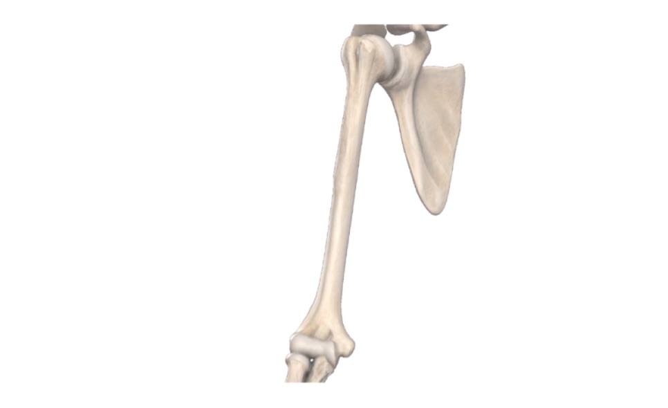
Humerus (Viva-related)
Q.1 How will you determine the side to which the humerus belongs?
- The upper end is rounded and forms the head. The lower end is flattened from before backward.
- Head is directed medially and backward.
- Lesser tubercle projects from the front of the upper end.
- The anterior aspect of the upper end shows a vertical groove called the intertubercular sulcus.
Q.2 What is the anatomical position of the humerus in the body?
- Head is directed medially, upwards and backward.
- The medial epicondyle is directed medially and slightly backward.
Q.3 What are the contents of the intertubercular sulcus (Bicipital groove)?
- The tendon of the long head of biceps.
- Synovial sheath of the tendon.
- Ascending branch of anterior circumflex humeral artery.
Q.4 What are the structures attached to intertubercular sulcus?
- To medial lip: Teres major, insertion.
- To floor: Latissimus dorsi, insertion.
- To lateral lip: Pectoralis major, insertion.
Q.5 How the tendon of the pectoralis major is inserted?
- It is inserted by a bilaminar tendon on the lateral lip of the bicipital groove, the two laminae are continuous with each other inferiorly.
Q.6 What are the muscles inserting on greater tubercle?
- Supraspinatus: On upper impression.
- Infraspinatus: On middle impression.
- Teres minor: On lower impression.
Q.7 Which muscle is inserted into lesser tubercle?
- Subscapularis
Q.8 What is the ‘anatomical neck’ and ‘surgical neck’ of the humerus?
- Anatomical neck: Surrounds the margin of head. Provides attachment to the capsular ligament which is deficient inferiorly on the medial side.
- Surgical neck: Lies at the upper end of the shaft, below the epiphyseal line. It is a common site for fracture.
Q.9 What are the structures related to the surgical neck of the humerus?
- Axillary nerve • Anterior and posterior circumflex humeral vessels
Q.10 Name the structures related to the radial groove of shaft of humerus.
- Radial nerve and
- Profunda brachii vessels
Q.11 Which muscle originates from medial supracondylar ridge?
- Pronator teres, from the lower end of the medial supracondylar ridge
Q.12 Which muscle originates from lateral supracondylar ridge?
- Brachioradialis: From upper two-thirds.
- Extensor carpi radialis longus: From lower one-third.
Q.13 Which muscles arise from the lateral epicondyle of the humerus (common extensor origin)?
- Extensor carpi radialis brevis
- Extensor digitorum
- Extensor digiti minimi
- Extensor carpi ulnaris
Q.14 Which muscles also arise from the lateral epicondyle of humerus except from common extensor origin?
- Anconeus
- Supinator
Q.15 What is the angle of ‘Humeral torsion’?
- It is an angle formed by the superimposition of long axes of upper and low articular surfaces of the humerus.
- It is about 164 degrees.
- It is greater in men and in adults.
Q.16 What are fossae present in the lower end of the humerus?
- Olecranon fossa: Posteriorly, just above trochlea. Accommodates the olecranon process in extended elbow.
- Radial fossa: Anteriorly, above capitulum. Accommodates radial head in flexed elbow.
- Coronoid fossa: Anteriorly, above trochlea. Accommodates coronoid process of ulna in flexed elbow.
Q.17 Why the fracture of the humerus at the junction of the upper and middle third leads to delayed union?
- Because of poor blood supply at the junction.
Q.18 Which nerve is most commonly involved in the supracondylar fracture of the humerus?
- Median nerve
Q.19 What are the ossification centers for the lower end of the humerus and at what age they appear?
- Capitulum: First year
- Medial epicondyle: Fifth year
- Medial part of trochlea: Ninth year
- Lateral epicondyle: Twelfth-year Medial epicondyle fuses with the shaft at 20th year and others fuse together to form a single epiphysis, which fuses with the shaft at 15 years of age.

Comments (0)