Radius
6 years ago 3391
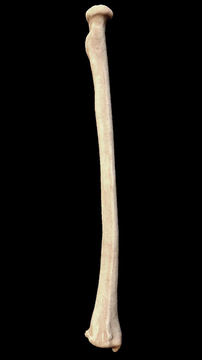
Parts of Radius
1. Upper End
• Head
• Neck
• Radial tuberosity
2. Lower end
3. Shaft
| Its shape is disc-like. |
| In living it is covered with articular hyaline cartilage. |
| During the full flexion, it articulates superiorly with the capitulum to form a humero-radial joint. |
| It articulates medially with the radial notch of the ulna. |
| All the parts are encircled by annular ligament except the articular part. |
Neck
→ It is the constricted part.
→ It lies below the head.
→ The quadrate ligament is attached to the side of the neck.
Radial tuberosity
It has two-part
• Smooth anterior part is covered by synovial bursa which separates this part from biceps tendon.
• The rough posterior part gives insertion to the biceps tendon.
It has three surfaces
|
It starts from the anterolateral part of the radial tuberosity.
|
| It runs downward and laterally to the styloid process. |
| It has two parts, the upper part (Anterior oblique line) and the lower part (Sharp). |
| Anterior oblique line Gives origin to the radial head of the flexor digitorum superficialis. |
Three-fourth parts of this border gives attachment to the interosseous membrane.
|
This is the area between the anterior and interosseous border.
|
| Nutrient foramen present in this surface which lies just above the middle part of the surface. |
| The direction of the nutrient canal is upward from where the nutrient artery passes which is a branch of the anterior interosseous artery. |
| Upper two-fourth Gives origin to the Flexor pollicis longus |
| Lower one-fourth Gives insertion to the Pronator Quardatus. |
| It is the area between the interosseous and posterior border. |
| Middle one-third Gives origin of the abductor pollicis longus |
| Lower part of this surface Give origin of the Extensor pollicis brevis |
|
It is the area between the anterior and posterior border.
|
| Upper one-third Give insertion to the supinator |
| Middle of the surface on the rough area Gives insertion to the pronator trees. |
| ¶ Anterior surface • A thick ridge is present on this surface provides attachment to the palmar radio-carpel ligament of the wrist joint. |
|
| ¶ Posterior surface • Here lister’s tubercle and dorsal tubercle of the lister present. |
|
| Dorsal tubercle of the lister • Lateral to this tubercle, a grove is present. • That is the groove for the extensor pollicis longus and other extensor tendons. |
|
| Lister’s tubercle • Lateral to this tubercle a grove is present which is transversed by the tendons of extensor carpi radialis longus and extensor carpi radialis brevis. • Medial to this grove extensor pollicis longus passes which is a common tendon of extensor digitorum and extensor indicis. |
|
| ¶ Medial surface • A notch called ulna notch is present on this surface. • It gives articulation to the head of the ulna and forms the radioulnar joint. • Articular disc of the inferior radio-ulnar joint is attached to the lower margin of the ulnar notch. Note:
|
|
| ¶ Lateral surface • It is projected downward as the styloid process. • It is related to the tendons of the abductor pollicis longus and extensor pollicis brevis. • The base of the styloid process gives insertion to the brachioradialis. • Tip of styloid gives attachment to the radial collateral ligament of the wrist joint. |
|
| ¶ Inferior surface It has two parts Lateral triangular area • Articulate with the scaphoid bone Medial quadrangular area • Articulate with lateral part of lunate bone |
Also read: Anatomy Question Collection
Also read: Anatomy Questions & Answers
Also read: Anatomy notes
Comments (0)