Humerus
6 years ago 3779
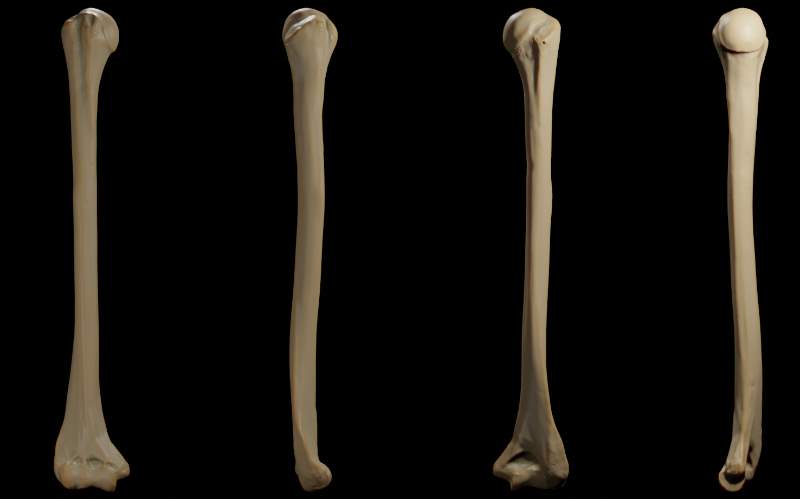
Parts of Humerus
1. Upper end
• Head
• Neck
• Greater tubercle
• Lesser tubercle
• Intertubercular sulcus
2. Lower end
- Articular part
• Capitulum
• Trochlea
- Non-articular part
• Coronoid fossa
• Radial fossa
• Olecranon fossa
3. Shaft
Upper end of Humerus
Head
Smooth and rounded.
Articular surface covered by hyaline cartilage.
Neck
There are three types of neck.
1. Anatomical neck
2. Surgical neck
3. Morphological neck
Anatomical neck
| It is the constriction present at the margin of the rounded head. |
| Gives attachment to the Capsular ligament of the shoulder joint. |
| Capsular ligament is deficient superiorly for the passage of the tendon of the long head of biceps brachii. |
| Capsular ligament extends medially from the anatomical neck to the shaft about 1-2 cm. |
Surgical neck
| It is the constriction present between the upper end of the shaft and below the greater and lesser tubercle. |
| It is related to the axillary nerve and posterior and anterior circumflex humeral vessels. |
| It is the weakest part of the humerus where fracture commonly takes place. |
| Fractures at this point lead to damage to the axillary nerve and vessel associated with it. That's why it is surgically important. |
Morphological neck
| It is the junction between epiphysis and diaphysis. |
| It is also the true junction of the head and shaft. |
Greater tubercle
| It is the most lateral part located at the proximal end of the humerus. |
| Posterio-superior parts bears three flat impression |
| Those flattened impressions give attachment to the supraspinatus, infraspinatus, and teres minor muscle to upper, middle, and lower impression respectively. |
Lesser tubercle
| Small elevation |
| Lie just above the surgical neck. |
| Give attachment to the subscapularis muscle. |
| Supraspinatus Infraspinatus Teres minor Subscapularis These are the muscles of rotator cuff |
Bicipital groove/ Intertubercular sulcus
| It is a vertical groove between lesser tubercle and greater tubercle. |
|
Content
|
| Lateral lip Gives attachment to the pectoralis major |
| Medial lip Gives attachment to the teres major |
| Floor of groove Gives attachment to the Latissimus dorsi |
Shaft of Humerus
Anterior border
| It begins from the lateral lip of the bicipital groove and runs anteriorly. |
| It continues below the margin of the deltoid tuberosity. |
| In the lower half it becomes smooth and rounded. |
| It ends in the radial fossa. |
Medial border
| It begins from the medial lip of the bicipital groove and gives continuous with the medial epicondyle. |
| Rough area in the middle of this border provides insertion to the coracobrachialis muscle. |
| Lower area of this border above the medial epicondyle gives origin to the pronator teres. |
Lateral border
| It is indistinct in the upper part but prominent in the lower part where it forms the lateral epicondyle. |
| Lower parts of this border gives attachment to the lateral intermuscular septum. |
Anterolateral surface
| It is the area between the anterior and lateral border. |
| Deltoid tuberosity present in this surface which is V-shaped. |
| Deltoid tuberosity is the main characteristics features of this surface. |
Anteromedial surface
| It is the area between the anterior and medial border. |
| Middle of this border nutrient foramen is present which is directed downward. |
Posterior surface
| It is the area between the medial and lateral border |
| An oblique ridge is present in the upper one-third of this surface that is directed downward and laterally. |
| Below the ridge there is the presence of a groove called radial groove which lodges radial nerve and profunda brachii vessels. |
| An oblique ridge Give origin to the lateral head of triceps brachii |
| Below the radial groove Gives origin to the medial head of triceps brachii |
Lower end of Humerus
Articular part:
Capitulum
• It is a rounded projection and lies laterally.
• Articulates with head of the radius.
Trochlea
• It is a pully shaped projection lies medially.
• Articulates with the trochlear notch of the ulna.
Non-articular part
Radial fossa
• Lies above capitulum.
• During the full flexion of the elbow joint, the head of the radius fits into the radial fossa.
Olecranon fossa
• Lies posterior aspect above the trochlea.
• During the full extension of the elbow joint, the olecranon process of the ulna fits into the olecranon fossa.
Coronoid fossa
• It lies above the trochlea.
• During full flexion of the elbow joint, the coronoid process of the ulna fits into coronoid fossa.
Attachment of lower end
| Anterior surface of the medial epicondyle Give the common origin of the superficial flexor of the forearm |
| Anterolateral part of the lateral epicondyle Give common extensor muscles origin |
| Posterior surface of the lateral epicondyle Gives origin to the anconeus muscle |
Note:
Relation of the nerve
| Axillary nerve → It around the surgical neck |
| Radial nerve → It passes through the radial groove |
| Ulnar nerve → It lies behind the medial epicondyle |
Also read: Anatomy Question Collection
Also read: Anatomy Questions & Answers
Also read: Anatomy notes
Comments (1)