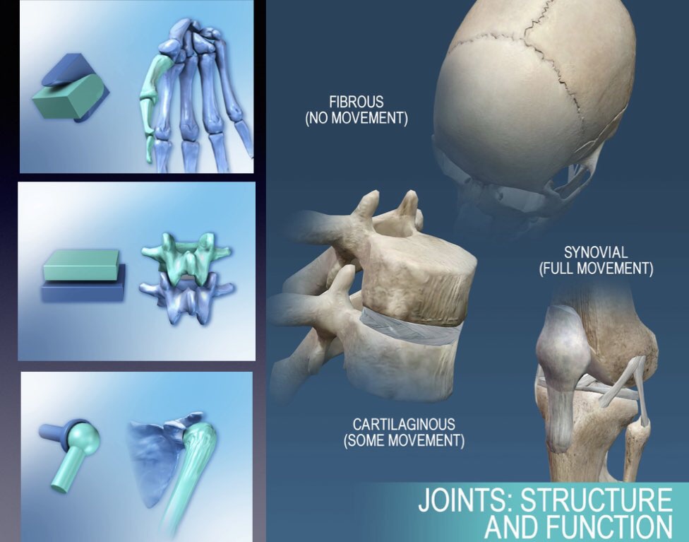
Joints Classification
6 years ago 8482

|
1. Synathroses
• Solid joints without any cavity • Example - Fibrous joints - Cartilaginous joints
|
|
2. Diarthroses
• Form synovial joints, which possess a joint cavity filled with synovial fluid and permit free movement • Example - Synovial joints |
| In these joints, bone are united by fibrous tissue. |
| Subdivided into - Suture Joint - Syndesmosis Joint - Gomphosis Joint |
| • Binding media is cartilage • Little movement possible |
|
Subdivided into
- Primary Cartilaginous (Synchondrosis) - Secondary Cartilaginous (Symphysis) |
| Cavity containing joint containing synovial fluid in it. |
|
Subdivided into
• Ball & Socket type of synovial joint • Saddle type of synovial joint • Condylar type of synovial joint • Ellipsoid type of synovial joint • Hinge type of synovial joint • Pivot type of synovial joint • Plane type of synovial joint |
| • All joints present in the skull is of Suture type of fibrous joint except - AtlantoOccipital Joint - Temperomandibular joint - Joint between Socket present in mandible & maxilla with teeth |
| • Uniting media is dense fibrous tissue |
| • The uniting fibrous tissue may be replaced by bones tissue in later life |
| 1. Serrate suture: • Saw tooth-like appearance • Example Sagittal suture of the skull |
|
2. Denticular suture:
• Teeth like margin • Eg.Lamboid suture
|
|
3. Squamous suture:
• Edges are united by overlapping
• Eg. Between parietal & squamous part of the temporal bone of the skull
|
| 4. Limbous suture: • Eg. Mastoid process of temporal bone |
|
5. Wedge & Groove suture:
• Eg.Between the rostrum of sphenoid and Vomer |
|
6. Plane suture:
• Border are plane • Eg.Between palatine processes of maxilla
|
| Binding media is Hyaline cartilage in nature |
| No movement |
| Temporary and the cartilages become ossified replaced by bone (called as synostosis) |
|
Example:
• Junction between epiphysis and diaphysis of growing long bone • 1st chondrosternal joint • Xiphisternal joint • Articulation between basi-occipital and basi-sphenoid (between basilar part of occipital bone & Body of Sphenoid bone)
|
| Synostosis Means bone to bone joint by bonny tissues |
|
Fate of Primary Cartilaginous Joint
With the age, the Cartilage is wholly replaced by complete bony Union i.e has formed synostosis |
| Articular surfaces of bones are covered with hyaline cartilage and are united by a plate of fibrocartilagenous structure (which is present between the middle of joint) |
| Permanent - persist throughout the life |
|
Example:
• All joint lying in the midline of body except xiphisternal joint
• Intervertebral joint between bodies of vertebrae • Symphysis menti • Pubic Symphysis • Sterno-manubrial joint • Lumbosacral joint • Sacrococcygeal joint |
| 1. Articular surface of bones are covered by hyaline cartilage |
| 2. Joint present a cavity which is filled with colorless synovial fluid |
| 3. Joint cavity is enveloped by an articular capsule, which consists of an outer fibrous capsule and inner synovial membrane |
| 4. The synovial membrane lines the whole of the interior of the joint cavity except the articular surface which is lined by hyaline cartilage. |
| 5. Sometimes, the joint cavity is divided into two compartments by fibrocartilagenous structures like articular disc, meniscus. Like in temporomandibular joint cavity has two compartments divided by articular disc. |
| 6. Movement is permitted from limited to a wide range |
| 7. Articulating bones are connected by a no.of accessory ligament |
| Four distinguishing features of synovial joint are: |
| • Joint cavity • An articular cartilage (Articular surface lined by hyaline cartilage) • A synovial membrane (which produce synovial fluid) • An articular capsule |
| Movement over one axis only Uniaxial movement i.e - Flexion & Extension |
|
Example
• Elbow joint (Humeroulnar-Humeroradial joint)
• Ankle Joint
• Interphalangeal joints of finger & toes |
|
Uniaxial movement i.e
- Flexion & extension
|
| Example • Proximal radioulnar joint • Distal Radioulnar joint • Median atlanto_axial joint |
|
Movement:
Flexion & Extension
|
| Example • Temperomandibular joint • Knee joint |
|
Movement over two axis
Biaxial movement: - Flexion, extension,
- Abduction, abduction,
- and Circumduction
|
| Example • Metatarsal-phalangeal joint • Metacarpal-phalangeal joint • Wrist joint (RadioCarpal joint) • AtlantoOccipital joint |
|
Example
• Sternoclavicular joint • 1st carpometacarpal • Between patella & femur |
|
Multiaxial movement:
- Flexion, extension,
- Abduction, abduction,
- Medial & lateral rotation,
- and circumduction
|
| Example • Hip joint • Shoulder joint • Between incus & stapes |
| Only gliding movement |
| Example • Sacroiliac joint • Costovertebral joint • Costotransverse joint • Between articular processes of vertebrae (facet joint) • Except 1st chondrosternal joint • Interchondral joint (5-9th) • Intertarsal joint • Intercarpal joint • All Carpometacarpal (except 1st) • Tarsometatarsal joint • Superior tibiofibular joint |
| - Possesses two articular surfaces - And only one joint cavity |
|
Example
• Hip joint • Interphalangeal joint of finger & toe
|
| - Possesses more than two articular surfaces - But only one joint cavity |
| Example • Wrist joint(radio-carpal joint) • Ankle joint |
| Possesses more than two articular surfaces |
| and two joint cavity, formed by the presence of articular disc or meniscus |
| Example • Knee joint • Sternoclavicular joint • Temperomandibular joint |
| When joint move around one axis only |
| Example |
| • Joints of Hinge type (elbow joint) | Flexion & extension |
| • Joints of Pivot type (Proximal radioulnar joint) | Rotation only |
| When joints moves around two axis |
| Example |
| • Joints of condylar type (knee joint) | Flexion, extension & limited rotation |
| • Joints of ellipsoid type (wrist joint) | Flexion, extension Abduction, adduction, circumduction |
| When joints move around more than two axis |
| Example |
| • Joints of saddle type(1st carpometacarpal joint) | Flexion, extension, Abduction, adduction & - In thumb Additional Opposition movement |
| • Joints of ball & socket type(hip joint) |
Comments (0)