Why Dental Implants Fail: Peri-Implantitis And The Diagnostic Gaps Most Patients Never Hear About
22 hours ago 35

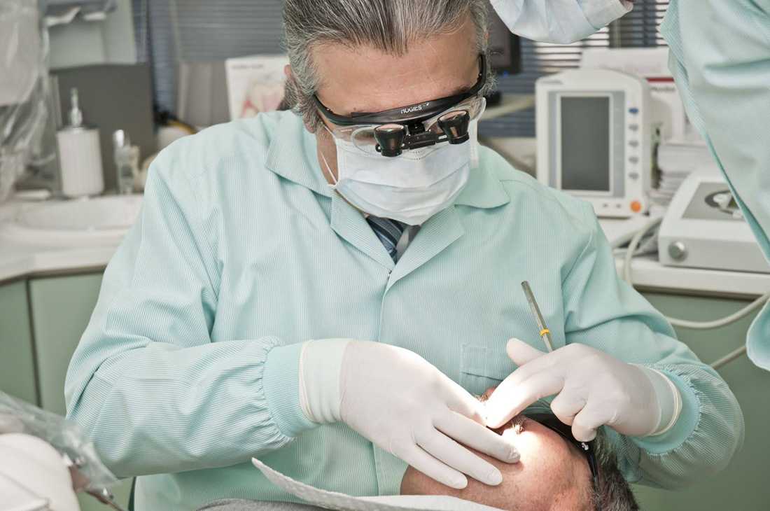
Dental implants get talked about almost exclusively in how successful they tend to be. That framing isn't wrong, but it's incomplete in a way that matters. Success rates describe outcomes at a population level. They don't explain what actually determines which side of that percentage a given patient lands on, and the answer has less to do with the implant itself than with a set of diagnostic and biological factors that rarely make it into consumer-facing content.
The single biggest driver of implant failure isn't a defective device or a technical slip during placement. It's a condition called peri-implantitis, and it's far more common and preventable than most patients realize.
Peri-implantitis is inflammatory bone loss around a dental implant, driven by a bacterial process similar to periodontal disease around natural teeth. It typically starts as peri-implant mucositis, inflammation confined to the soft tissue, and progresses to peri-implantitis once the underlying bone begins to resorb. Left unaddressed, that bone loss is what ultimately causes implants to loosen and fail.
The prevalence numbers are wider-ranging than most people expect. A retrospective study of electronic health records at a U.S. dental school, covering over 6,000 implants, found peri-implantitis in 34 percent of patients and 21 percent of individual implants over an average two-year follow-up.
This reflects real, identifiable differences in patient risk factors and, notably, in the diagnostic and surgical rigor behind how the implant was placed in the first place.
A few factors show up consistently across the research as the strongest predictors of trouble down the road.
Adequate bone quantity is a genuine prerequisite for stable, successful restoration. Patients with bone deficiencies at the surgical site face measurably higher risk of poor implant stability, delayed osseointegration, and bone remodeling failure. In simple terms: if there isn't enough healthy bone in the right place before the implant goes in, the odds of a problem later go up substantially, regardless of how well the implant itself is manufactured.
The issue is that this kind of deficiency isn't always visible on a standard exam or a flat two-dimensional X-ray. It requires volumetric imaging, most commonly cone-beam CT, to actually measure bone density and quantity in three dimensions before a surgical plan is finalized.
That step is what allows a provider to catch a deficiency in advance and address it through bone grafting or guided regeneration before placing the implant, rather than discovering the problem after the fact when the implant is already failing.
This is precisely where training differences among providers start to matter in a very concrete way. Diagnosing and correcting bone volume deficiencies, through ridge augmentation, sinus lifts, or guided bone regeneration, sits squarely within the surgical training of oral and maxillofacial surgeons, whose residency programs run several years beyond dental school and include the kind of hospital-based surgical training that general dentistry programs don't require.
A provider equipped to diagnose and correct bone volume issues before placement, not after who can identify a bone deficiency and correct it in the same treatment plan is working from a fundamentally different risk profile than one who places implants without that surgical depth to fall back on.
None of this is meant to suggest implants are a risky choice. The overall success rates remain high, and for the large majority of patients, a well-planned implant is a genuinely durable, low-maintenance solution to tooth loss.
The point is narrower: failure, when it happens, is rarely random. It tends to trace back to one of a small number of identifiable factors, untreated periodontal history, uncontrolled diabetes or smoking, inconsistent maintenance, or a bone deficiency that wasn't properly assessed before surgery.
Of those, the last one is the most preventable, because it's entirely a function of the diagnostic process before the first incision, not something the patient has much control over once treatment has begun.
Asking a provider directly about their imaging capabilities, how they assess bone volume, and what their plan is if a deficiency turns up is a reasonable, non-confrontational way to understand which side of the statistics a given treatment plan is more likely to land on.
Comments (0)