Inclusion bodies
Inclusion bodies are dense, insoluble aggregates of proteins or other biomolecules such as nucleic acid that accumulate inside the cells. Inclusion bodies can be either cytoplasmic or nuclear based on their location and can be observed under a microscope.
Formation of Inclusion bodies
Typically, Inclusion bodies are dense, insoluble aggregates of proteins often form as a result of cellular stress, overexpression of recombinant proteins or protein misfolding. There are several explanation about inclusion bodies formation, like:
- Protein misfolding: When synthesis of proteins occur at high rates, they may not fold correctly and can form aggregates. These misfolded proteins can then accumulate and form inclusion bodies.
- Incomplete protein degradation: Sometimes, proteins are not properly degraded byproteasome, a cellular complex that breaks down proteinsand accumulate forming inclusion bodies.
- Overexpression of recombinant proteins: When a foreign protein is introduced into a cell, it may be overexpressed, leading to the formation of inclusion bodies. When genes from one organism are expressed in another organism, such as a cDNA from eukaryotes in a prokaryote, inclusion bodies may form. The microenvironment of the host organism may differ from that of the source of the gene, and necessary mechanisms for folding, processing, and concentration control may be absent, leading to inclusion bodies formation.
- Cellular stress: Stressful conditions, such asexposure to chemicals orhigh temperature, can cause proteins to misfold and aggregate, resulting in inclusion bodies formation.
Overall, the formation of inclusion bodies is a complex process that can be influenced by a variety of factors, including protein structure, expression levels, and cellular environment.
Classification of Inclusion Bodies
Inclusion bodies are found in number of tissue cells like neurons, red blood cells, white blood cells, bacterial infection, virus infected cells, plant cells, and muscle cells affected by inclusion body myositis and hereditary inclusion body myopathy. Different types of inclusion bodies are:
- Intracytoplasmic inclusion bodies
- Intranuclear inclusion bodies
- Physiological inclusion bodies
- Infection inclusion bodies (Bacterial, Viral, Fungal Infection)
- Inclusion bodies in Neoplasm
- Inclusion bodies in Autoimmune diseases
Inclusion bodies found in Neuron
In neurons, presence of inclusion bodies can be a hallmark diagnostic clue of various neurodegenerative diseases, such as Alzheimer's disease, Parkinson's disease, and Huntington's disease.
- Lewy bodies
- Neurofibrillary tangles
- Huntingtin aggregates
- Inclusion body myositis
Lewy bodies: Lewy bodies are spherical inclusion bodies found in the neurons of people with Parkinson's disease and Lewy body dementia. Lewy bodies is the aggregation of alpha-synuclein protein.
Neurofibrillary tangles: Neurofibrillary tangles are the inclusion bodies found inside the brain cells of people with Alzheimer's disease. Neurofibrillary tangles are twisted fibers that are composed of a protein called tau, which normally helps stabilize the structure of nerve cells.
Huntingtin aggregates: In Huntington's disease, huntingtin protein aggregates as intracellular inclusion bodies called huntingtin aggregates. These aggregates lead to progressive neuronal death and the symptoms associated with the disease.
Inclusion body myositis: Inclusion body myositis (IBM) is a rare progressive muscle disorder that primarily affects older adults, characterized by the presence of inclusion bodies within muscle cells, which are abnormal clumps of protein that accumulate in the cytoplasm. IBM is considered to be both a myopathy (muscle disease) and a neuropathy (nerve disease).
Inclusion bodies found in RBC
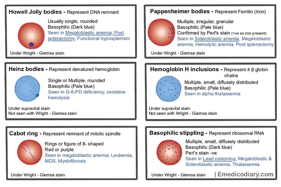
Generally, RBCs have no nucleus or organelles and inclusion bodies are not present in RBCs. However, there are a some conditions where inclusion bodies can be present in RBC.
Inclusion bodies found in RBC are
- Basophilic stippling
- Pappenheimer bodies
- Howell Jolley bodies
- Heinz bodies
- Hemoglobin H inclusion
- Cabot rings
- Protozoan inclusion in Malaria, Babesia
Basophilic stippling (Punctate basophilia):
Basophilic stippling also known as punctate basophilia are small, blue black inclusion bodies scattered throughout the cytoplasm of RBCs and represent precipitated ribosomal RNA. They do not stain with Perl's stain (i.e. no iron present in Basophilic stippling).
Basophilic stippling is found in Lead poisoning, Megaloblastic anemia, Thalassemia, Sideroblastic anemia and alcoholism.
Pappenheimer bodies
Pappenheimer bodies represents aggregation of ferritin (Iron) in RBCs and appears as pale blue, fine and irregular granules at the periphery of the RBCs. They are easily demonstrable with Perl's stain (Prussian blue reaction) as they contain iron. They are also known as siderotic granules. RBCs with Pappenheimer bodies are called siderocytes.
Pappenheimer bodies are found inSideroblastic anemia, Megaloblastic anemia, Hemolytic anemia, Post splenectomy.
Howell Jolley bodies
Howell Jolley bodies are the small, round basophilic (dark blue) inclusion bodies found in RBCs and represent nuclear remnants (DNA fragments). RBCs lost their nucleus during their maturation process in bone marrow, but when RBCs fails to maturate properly, nuclear remnants persists and appears as dense basophilic granule known as Howell Jolley bodies.
Howell Jolley bodies are commonly found in Megaloblastic anemia, Post splenectomy, Functional hyposplenism. Splenectomy is done as therapeutic purposes in Hereditary spherocytosis, spleen trauma, and auto-splenectomy caused by sickle cell anemia. Functional hyposplenism seen in sickle cell anaemia, splenic artery or vein thrombosis, SLE, liver cirrhosis, amyloidosis, sarcoidosis, etc.
Heinz bodies
Heinz bodies are refractive, single or multiple rounded inclusion present in RBCs composed of denatured hemoglobin. They are not stained by routine hematological stain such as Wright - Giemsa stain. They are visible by Supravital stain.
Heinz bodies are found in G-6-PD deficiency, oxidative hemolysis due to intake/exposure to oxidizing drugs or chemical.
Cabot Rings
Cabot rings are the rings or figure of eight shaped structure present in RBCs and are probably remnants of mitotic spindle. Cabot Rings are found in megaloblastic anemia, Leukemia, MDS, Myelofibrosis.
Hemoglobin H inclusion
Hemoglobin H inclusions are free β chains in red cells appears as small, multiple and diffusely distributed. They are stained by supravital stains. Hemoglobin H inclusions are found in alpha-thalassemia.
Malarial Pigments
Plasmodium feeds on hemoglobin. The undigested product of hemoglobin metabolism like hematin, excess protein and iron porphyrin combine to form malarial pigment called hemozoin.
Inclusion bodies in WBC
Several inclusion bodies can be found in white blood cells like
- Dohle bodies
- Auer rods
- Russell bodies
Dohle bodies: Dohle bodies are small, round, blue-gray, basophilic inclusion bodies found in the cytoplasm of neutrophils. They are thought to be caused by the aggregation of ribosomal RNA and rough endoplasmic reticulum, and can be seen in certain bacterial infections, burn, inflammatory conditions, and after chemotherapy.
Auer rods: Auer are rod-shaped, eosinophilic crystalline inclusion bodies found in the cytoplasm of myeloblasts (an immature type of WBC) in patients with acute myeloid leukemia. Auer rods are composed of a protein called myeloperoxidase and are considered to be a diagnostic feature of this type of leukemia.
Russell bodies: Russell bodies are eosinophilic, round or oval-shaped inclusion bodies that are found within the cytoplasm of plasma cells. These structures are composed of immunoglobulins that have accumulated in the endoplasmic reticulum of the plasma cells. Russell bodies are commonly associated with multiple myeloma, a type of blood cancer that involves the uncontrolled growth of plasma cells. They can also be seen in other types of B-cell neoplasms and in certain inflammatory conditions.
Viral Inclusion bodies
Viral inclusion bodies are aggregates of virus particles or virus-induced proteins that form inside infected host cell either in cytoplasm, nucleus or both. The presence of inclusion bodies can be used as a diagnostic tool for identifying viral infections. Different viruses may produce distinct inclusion bodies, which can aid in identifying the specific virus responsible for the infection. Some examples of viral inclusion bodies include:
A. Intracytoplasmic eosinophilic inclusion bodies
- Negri bodies in neuron in rabies virus
- Guarnieri bodies in vaccinia, variola (smallpox)
- Paschen bodies in variola (smallpox)
- Bollinger bodies in fowlpox
- Molluscum bodies or Handerson-Patterson bodies in Molluscum contagiosum
- Eosinophilic inclusion bodies in boid inclusion body disease
- Councilman Bodies found in cytoplasm of hepatocytes, with ballooning degeneration in viral infection like viral hepatitis, yellow fever
B. Intranuclear eosinophilic
- Torres bodies in yellow fever
- Cowdry type A in Herpes simplex virus & Varicella zoster virus (Chickenpox)
- Cowdry type B in Polio
C. Intranuclear basophilic
- Cowdry type B in Adenovirus
- Owl's eye inclusion bodies in cytomegalovirus
D. Both nuclear and cytoplasmic
- Warthin–Finkeldey bodies in measles
- Many small inclusion bodies in cytoplasm and intranuclear owl's eye inclusion bodies in cytomegalovirus
Inclusion bodies in neoplasm
- Psammoma bodies are esosinophilic, concentrically laminated, calcified mass formed due to necrosis followed by dystrophic calcification. Psammoma bodies seen in Papillary thyroid carcinoma, Papillary renal cell carcinoma, Papillary serous cystadenocarcinoma of ovary, Endometrial adenocarcinomas, Meningiomas,Peritoneal and Pleural Mesothelioma
- Russell bodies are seen in multiple myeloma, plasmacytoma
- Dutcher bodies seen in chronic synovitis and large B- cell lymphoma and multiple myeloma.
- Pustulo- Ovoid bodies seen in granular cell tumors
- Verocay bodies seen in Schwannoma
- Kamino bodies seen in pigmented spindle cell nevus, Spitz nevus
- Wagner- Meissner body seen in von Recklinghausen’s disease of skin, neurofibroma
Inclusion Bodies in autoimmune diseases
- Asteroid bodies are star-shaped inclusion bodies with numerous rays radiating from the central core found in sarcoidosis, sporotrichosis (fungal infection).
- Civatte bodies seen in discoid lupus erythematosus and lichen planus
- Hematoxylin bodies seen in systemic lupus erythematous
- Schaumann bodies seen in Sarcoidosis, tuberculosis, hypersensitive pneumonitis
Some other pathological named bodies
- Halberstaedter - Prowazek bodies seen inTrachoma
- Miyagawa bodies seen in Lymphogranuloma venereum
- Mooser bodies seen in Typhus bodies
- Levinthal Cole Lillie bodies seen in Psittacosis
- Leishman Donovan bodies seen in Kala-azar
- Donovan bodies seen in Granuloma Inguinale
- Creola bodies seen in Asthma
- Aschoff bodies seen in Rheumatic fever
- Verocay bodies seen in Schwannoma
- Hirano bodies seen in Alzheimer's diseases
- Lewy bodies seen in Parkinson's disease

Comments (0)