How Secondary Antibodies Improve IF & Flow Cytometry Precision
1 year ago 1385
Immunofluorescence microscopy and flow cytometry are two techniques that have become indispensable for visualizing protein localization and quantifying cell-surface markers.
While these two may sound complicated, both methods use a deceptively simple tool, which is the secondary antibody.
Let’s explore how a well-chosen secondary antibody portfolio amplifies signal, refines specificity, and ultimately determines the clarity of experimental conclusions.
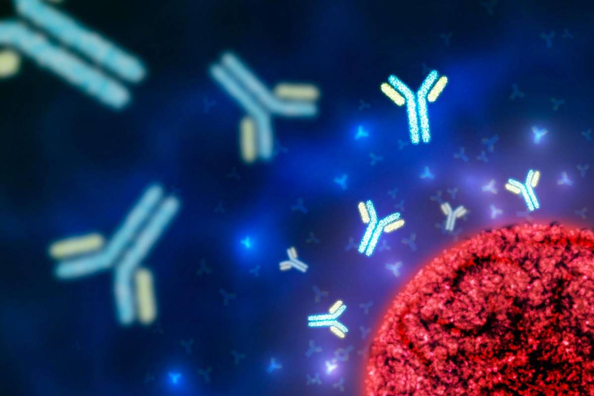
Primary antibodies bind directly to target antigens. Secondary antibodies, in turn, recognize the conserved regions (Fc domains) of those primaries.
The system multiplies the detectable signal by conjugating a detectable label, fluorophores, enzymes, or heavy-metal isotopes to the secondary antibody without altering the primary’s binding affinity. This modular two-step design offers three important benefits:
Under a fluorescence microscope, subcellular regions turn into glowing maps.
Photostable dyes like Alexa Fluor 488 or 594 guard against bleaching during prolonged exposure, while far-red options like Alexa Fluor 647 unlock super-resolution methods (STED, STORM) that push past the diffraction limit.
Species-specific secondaries make multiplexing straightforward: goat anti-mouse for one antigen, donkey anti-rabbit for another, and so on. Each channel retains its own spectral lane, ensuring accurate co-localization studies.
| Factors | Why It Matters |
| Fluorophore brightness | Supports lower laser power and shorter exposure |
| Photostability | Maintains image quality during z-stacks |
| Cross-adsorption level | Reduces tissue background |
| Aggregate content | Prevents artifactual puncta |
Flow cytometers count thousands of cells per second, measuring fluorescence intensity for each. Secondary antibodies labeled with tandem dyes (PE-Cy7, APC-H7) deliver the quantum yield needed to resolve dim antigens without raising laser power. Because isotype-matched primaries from different species can be paired with distinct secondaries, panels grow quickly, ideal for immunophenotyping or cell-cycle profiling.
High Quantum Yield – Bright dyes expose low-density markers.
Minimal Spill-Over – Properly chosen fluorochromes ease compensation.
Aggregate-Free Preparation – Smooth hydrodynamics prevent clogging and false doublets.
A comparative flow cytometry study assessed direct versus secondary-amplified detection of phospho-ERK. Directly labeled mouse anti-phospho-ERK yielded a mean fluorescence intensity (MFI) of 1,200. When the same primary was followed by goat anti-mouse IgG-PE, MFI jumped to 7,800—over sixfold improvement—while the coefficient of variation dropped below 5 %. Better separation between resting and activated cells demonstrated how secondary amplification strengthens biological conclusions.
Success in immunofluorescence and flow cytometry depends on more than advanced optics or complex software; it begins with a strategic choice of reagents. By supplying robust signal amplification, impeccable specificity, and broad versatility, secondary antibodies remain central to the accurate interpretation of protein expression. A disciplined selection process transforms these quiet helpers into powerful allies, illuminating cellular mysteries with clarity and confidence.
Comments (0)