Early Signals of Bone Loss and How to Take Action
1 year ago 512
Your skeleton undergoes continuous renewal through a careful equilibrium of bone dissolution and formation. When this balance shifts toward excessive breakdown, bone loss accelerates. This results in osteoporosis, which impacts more than 200 million individuals globally. The difficulty centers on identifying this degradation before a catastrophic break compels medical attention.
Detecting minor bodily changes can expose bone density loss years ahead of reaching dangerous levels, enabling intervention when your skeletal system remains highly responsive to protective treatments.
Measuring your height might seem like something you stopped doing after childhood, but tracking this simple metric could save you from debilitating fractures. Adults who lose more than one and a half inches from their peak height are experiencing vertebral compression, a clear indicator of weakening bones. These tiny fractures in the spine occur gradually, often without pain initially, as vertebrae slowly collapse under normal body weight.
The visual manifestation appears as a forward curve in the upper back, medically termed kyphosis or commonly called a dowager's hump. This isn't merely cosmetic; it represents structural failure in your spine. Each compressed vertebra reduces lung capacity, affects balance, and increases fall risk. Women may notice their ribs moving closer to their hip bones, causing the abdomen to protrude despite no weight gain.
Regular height measurements every six months after age 50 can catch this process early. Stand against a wall in the morning when spinal discs are fully hydrated for consistent measurements.
Document these numbers and report any loss exceeding half an inch per year to your healthcare provider. Incorporate spine-extending exercises like swimming backstroke and yoga poses such as cobra and bridge to counteract compression forces.
Sustaining a bone fracture from minimal force indicates advanced bone deterioration. Fragility fractures, from falls at standing height or less, indicate bones have lost significant density and structural integrity. Typical locations include wrists during falls, vertebrae when lifting modest weights, and ribs during forceful coughing.
These fractures frequently shock otherwise healthy, active individuals. The precipitating incident seems insufficient to cause fracture, yet imaging confirms clear breaks. Monitoring your bone healing timeline becomes essential, as delayed healing indicates persistent bone quality deficiencies requiring immediate intervention.
Hip fractures carry the most severe outcomes: 20% mortality within the first year, with only half of survivors recovering previous mobility. Even minor fractures warrant serious attention. Each fragility fracture doubles subsequent fracture risk, potentially triggering sequential skeletal failures without aggressive intervention combining lifestyle changes and medical treatment.
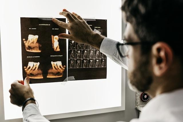
Your oral cavity provides a revealing window into your overall skeletal condition. The jaw, continuously regenerating like other bones, is equally susceptible to systemic bone deterioration. When the supporting alveolar bone weakens, gum recession occurs, teeth become unstable, and ultimately fall out, despite meticulous dental care.
Dental radiographs can detect declining jaw bone density before symptoms manifest elsewhere in the body. Research shows women with osteoporosis have three times higher tooth loss than those with normal bone density. Men experience similar patterns of oral bone deterioration, though typically appearing about 10 years later.
Be alert to newly loosened teeth, increasing spaces between teeth, or receding gums. Ill-fitting dentures that previously fit properly suggest ongoing jawbone loss. These indicators merit evaluation by dental professionals and comprehensive skeletal density assessment, as jaw bone deterioration frequently precedes hip and vertebral degradation by several years.
Your grip strength reveals bone health status, especially in postmenopausal women. Research shows direct links between weakening grip and declining bone mineral density throughout the skeleton. Both conditions share common causes: hormonal shifts, nutritional deficiencies, and physical inactivity.
Watch for difficulty opening jars, increased object dropping, or struggling with grocery bags. These signals indicate potential bone deterioration, not just normal aging. Dynamometer testing provides objective measurement, with readings below 20 kg for women and 30 kg for men indicating heightened fracture risk.
Grip strength effectively responds to intervention. Regular resistance training with hand grippers, stress balls, and elastic bands improves grip capability and bone density. Begin with manageable resistance, gradually increasing intensity across three weekly sessions. This approach simultaneously enhances daily functional capacity and stimulates systemic bone formation.
Perhaps most concerning is experiencing any of these symptoms before traditional risk periods, menopause for women, or age 70 for men. Early-onset bone loss suggests underlying conditions requiring immediate investigation: hormonal imbalances, malabsorption disorders, genetic predispositions, or medication effects.
Young adults with stress fractures, women under 50 with fragility fractures, or men under 70 with unexplained height loss need comprehensive evaluation beyond standard bone density testing. Investigation should include hormone panels, celiac testing, vitamin levels, and review of all medications. Common culprits include long-term proton pump inhibitors, certain antidepressants, and corticosteroids.
Fingernails provide visible indicators of your mineral status and corresponding bone condition. Chronic brittleness, vertical ridges, frequent splitting, or delayed growth may signal inadequate nutrient delivery to both nails and skeletal tissues. The minerals crucial for bone matrix development are:
Inspect your nails under adequate lighting, monitoring any consistent changes across multiple months. Healthy nails exhibit smooth surfaces, gentle curvature, and normal pressure resistance. White spots, pronounced ridges, or layered peeling suggest mineral inadequacies likely affecting your entire skeletal framework.
Mid to lower spine pain that worsens when standing may indicate vertebral compression fractures. Unlike acute injuries, this pain develops gradually. Distinctive pain patterns aid diagnosis: relief when recumbent, specific vertebral tenderness, and pain radiating anteriorly around the ribs. Patients often describe sensations of spinal collapse or carrying excessive back weight. These perceptions accurately reflect the mechanical failure of vertebrae that cannot bear normal loads.
Diagnostic imaging may show wedge-shaped vertebrae, reduced disc spaces, or evident compression fractures. However, early-stage bone deterioration causes pain before structural changes appear radiographically. Don't dismiss persistent back pain as normal aging; seek a comprehensive evaluation, including bone density assessment and metabolic testing, to identify the correct causes.
Identifying early-onset bone loss offers the best prognosis, as younger bones respond more robustly to intervention. However, it requires vigilance and advocacy, as many healthcare providers don't expect bone problems in younger patients. Document symptoms carefully, request appropriate testing, and don't accept dismissive responses to legitimate concerns about your skeletal health.
Comments (0)