How Researchers Visualize Complex Biological Structures
1 year ago 900

Understanding life at its tiniest levels—think cells, proteins, and DNA—starts with being able to see it. For researchers, visualizing these biological structures isn’t just a neat trick; it’s the key to figuring out what they do and how they work together. Whether it’s unraveling the mysteries of a disease or designing a new drug, seeing these intricate systems up close drives progress in medicine and science. Biological structures are anything but simple, so scientists have come up with some pretty clever ways to study them.
The demand for sharper, more detailed images has exploded lately, thanks to cutting-edge imaging tools that let us peek into the molecular world like never before. Being able to zoom in to the atomic level helps pinpoint what goes wrong in diseases, craft better treatments, and even develop things like vaccines. In this piece, we’ll dive into the techniques researchers use to make these hidden worlds visible and explore why those images matter so much to science.
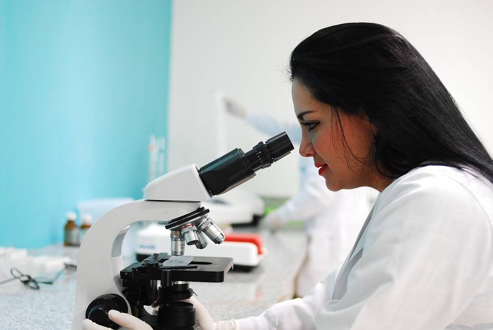
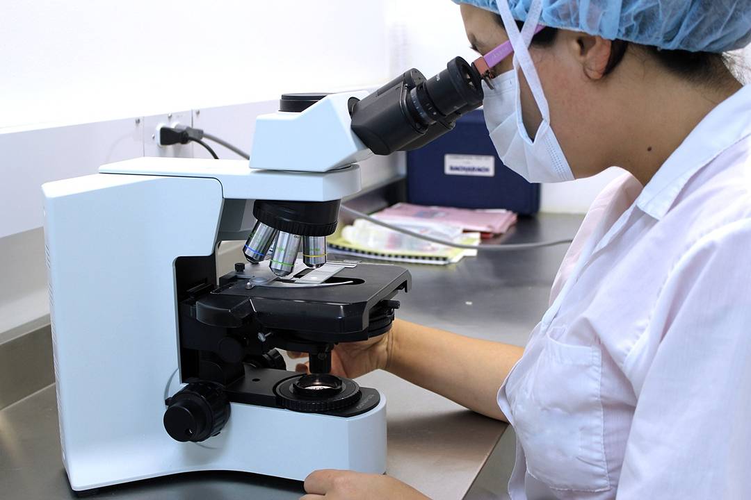
Let’s start with a tried-and-true method: light microscopy. It’s been around forever and uses basic visible light and lenses to blow up tiny objects for a closer look. Scientists rely on it to check out cells and tissues—things like the nucleus, mitochondria, or cytoskeleton pop into view. It’s great for getting the big picture, but there’s a catch. The resolution tops out around 200 nanometers, so forget about seeing individual molecules or the really fine details.
Now, fluorescence microscopy takes things up a notch. By tagging specific molecules with glowing dyes, it lights up the stuff scientists want to track. Shine the right wavelength of light on the sample, and those dyes glow back at a different color, making it easy to spot them. The use of a fluorescence microscope is a game-changer for watching live cells in action—think proteins moving around or signals firing off. It’s like giving researchers a front-row seat to biology in real time.
Confocal microscopy steps it up even further. It uses a laser to zero in on one tiny spot at a time and a clever pinhole to block out blurry light. The result? Crisp, clear images. Plus, it can stack those images into 3D models, which is perfect for digging into thick samples like tissues or embryos. When you need depth and precision, this is the go-to.
Then there’s super-resolution microscopy, which basically says, “Limits? What limits?” Methods such as STORM and PALM break through the former limits of diffraction of lights, allowing researchers to observe objects as small as 100 nanometers or even smaller. Such a size is tiny enough to witness molecular apparatus; for instance, ribosomes or complex proteins, in operation. This represents enormous progress when it comes to exploring the smallest foundation units of life.
Switching gears to electrons, scanning electron microscopy blasts a sample’s surface with a beam to create stunning 3D images. It’s way more powerful than light microscopy and can zoom in on things like viruses or cell surfaces with incredible detail. You’ll see it a lot in microbiology or materials science. The downside? Samples often need a metal coating, which can mess with their natural state a bit.
Transmission electron microscopy takes a different approach. Instead of scanning the surface, it shoots electrons straight through a super-thin slice of the sample. The payoff is jaw-dropping resolution—down to the atomic level. It’s perfect for exploring the insides of cells, viruses, or big molecular complexes. But it’s not easy: samples have to be sliced razor-thin and prepped just right to hold up under the electron barrage.
Cryo-electron microscopy is the new kid on the block, and it’s a total game-changer. By flash-freezing samples, it locks their natural structure in place—no crystals needed. That means researchers can study proteins and other molecules as they really are, not some altered version. Cryo-EM has cracked open the secrets of tricky protein structures, fueling breakthroughs in drugs and our grasp of how cells tick.
X-ray crystallography is a classic in the structural biology world. It works by firing X-rays at a crystalized sample and reading the patterns they scatter. From there, scientists piece together a 3D atomic map. It’s delivered huge wins—like decoding DNA or enzymes—but growing those perfect crystals can be a headache, especially for big, floppy proteins.
Nuclear magnetic resonance spectroscopy skips the crystal step entirely. It uses magnetic fields to map out a molecule’s atoms while it’s floating in solution, giving a peek at how it moves and flexes. That’s a big deal for studying proteins or DNA in their natural state. The catch? It struggles with really massive molecules, which can muddy the signal.
AI and machine learning are shaking things up big time. They’re sifting through mountains of data from tools like Cryo-EM to build sharper, faster models of biological structures. AI can even predict how molecules might interact, saving researchers tons of guesswork. It’s like having a super-smart assistant that speeds up the whole process.
And here’s something cool: virtual and augmented reality. Scientists can now step inside 3D models of cells or molecules, poking around like they’re in a sci-fi movie. It’s not just fun—it’s a powerful way to explore complex data. For students or researchers, VR and AR make learning and discovery way more hands-on.
Using tools like microscopes and computer models for visualizing biological structures, we are finding new ways to comprehend life more profoundly. These instruments are not just presenting us with attractive images, but they're propelling the development of new medicines, therapies and comprehensions that will mold the imminent era. Whether it’s zooming into a protein or walking through a cell in VR, this is science at its most exciting.
Comments (0)